Explore
 Validate
 Learn
Learn12-1548-41
antibody from Invitrogen Antibodies
Targeting: CD40LG
CD154, CD40L, gp39, hCD40L, HIGM1, IMD3, TNFSF5, TRAP
 Flow cytometry
Antibody data
- Antibody Data
- Antigen structure
- References [3]
- Comments [0]
- Validations
- Flow cytometry [1]
Submit
Validation data
Reference
Comment
Report error
- Product number
- 12-1548-41 - Provider product page

- Provider
- Invitrogen Antibodies
- Product name
- Anti-CD154 (CD40 Ligand) Monoclonal Antibody (24-31), PE, eBioscience™
- Antibody type
- Monoclonal
- Antigen
- Other
- Description
- Description: The 24-31 monoclonal antibody reacts with human CD154, a 39 kDa transmembrane glycoprotein also known as gp39 and CD40 ligand (CD40L). CD154 is a member of the TNF superfamily and is expressed transiently by activated T cells. Through its binding to CD40 on antigen presenting cells including B cells, monocytes/macrophages and dendritic cells, CD154 serves a crucial function in T-APC cognate interaction. CD154 interaction with CD40 transduces signals for T-dependent B-cell activation and induces B cell cycle entry. 24-31 cross-reacts with the cynomolgus monkey CD154. Applications Reported: 24-31 has been reported for use in flow cytometric analysis. Applications Tested: This 24-31 antibody has been pre-titrated and tested by flow cytometric analysis of activated normal human peripheral blood cells. This can be used at 5 µL (0.5 µg) per test. A test is defined as the amount (µg) of antibody that will stain a cell sample in a final volume of 100 µL. Cell number should be determined empirically but can range from 10^5 to 10^8 cells/test. Excitation: 488-561 nm; Emission: 578 nm; Laser: Blue Laser, Green Laser, Yellow-Green Laser. Filtration: 0.2 µm post-manufacturing filtered.
- Reactivity
- Human
- Host
- Mouse
- Conjugate
- Yellow dye
- Isotype
- IgG
- Antibody clone number
- 24-31
- Vial size
- 25 Tests
- Concentration
- 5 µL/Test
- Storage
- 4° C, store in dark, DO NOT FREEZE!
Submitted references Photodynamic therapy reduces the inhibitory effect of osteosarcoma cells on dendritic cells by upregulating HSP70.
Isolation of biologically-active exosomes from human plasma.
Endothelial microparticles interact with and support the proliferation of T cells.
Zhang F, Zhu Y, Fan G, Hu S
Oncology letters 2018 Oct;16(4):5034-5040
Oncology letters 2018 Oct;16(4):5034-5040
Isolation of biologically-active exosomes from human plasma.
Muller L, Hong CS, Stolz DB, Watkins SC, Whiteside TL
Journal of immunological methods 2014 Sep;411:55-65
Journal of immunological methods 2014 Sep;411:55-65
Endothelial microparticles interact with and support the proliferation of T cells.
Wheway J, Latham SL, Combes V, Grau GE
Journal of immunology (Baltimore, Md. : 1950) 2014 Oct 1;193(7):3378-87
Journal of immunology (Baltimore, Md. : 1950) 2014 Oct 1;193(7):3378-87
No comments: Submit comment
Supportive validation
- Submitted by
- Invitrogen Antibodies (provider)
- Main image

- Experimental details
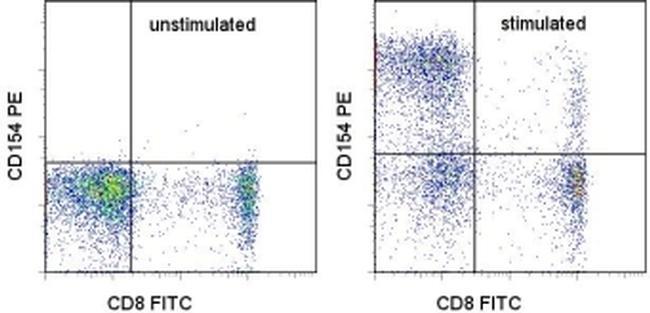
- Human peripheral blood cells unstimulated (left) or stimulated for 5 hours with Cell Stimulation Cocktail (500X) (Product # 00-4970-03) (right) were stained with Anti-Human CD8a FITC (Product # 11-0088-42) and Anti-Human CD154 (CD40 Ligand) PE (right). Cells in the lymphocyte gate were used for analysis.
- Conjugate
- Yellow dye