 Explore
 Validate
 Learn
Learn Western blot
Antibody data
- Antibody Data
- Antigen structure
- References [0]
- Comments [0]
- Validations
- Western blot [3]
- Immunocytochemistry [1]
- Immunohistochemistry [4]
Submit
Validation data
Reference
Comment
Report error
- Product number
- GTX101648 - Provider product page

- Provider
- GeneTex
- Proper citation
- GeneTex Cat#GTX101648, RRID:AB_1949564
- Product name
- AChE antibody
- Antibody type
- Polyclonal
- Reactivity
- Human, Mouse, Rat
- Host
- Rabbit
No comments: Submit comment
Supportive validation
- Submitted by
- GeneTex (provider)
- Main image
- Experimental details
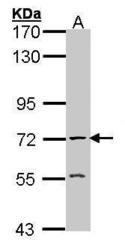
- Sample (30 ?g of whole cell lysate) A: Raji 7.5% SDS PAGE GTX101648 diluted at 1:1000 The HRP-conjugated anti-rabbit IgG antibody (GTX213110-01) was used to detect the primary antibody.

- Submitted by
- GeneTex (provider)
- Main image
- Experimental details
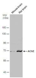
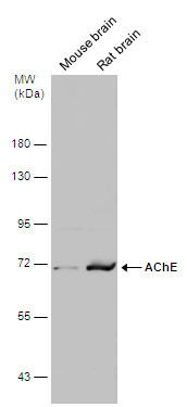
- Various tissue extracts (50 ?g) were separated by 7.5% SDS-PAGE, and the membrane was blotted with AChE antibody (GTX101648) diluted at 1:500. The HRP-conjugated anti-rabbit IgG antibody (GTX213110-01) was used to detect the primary antibody.
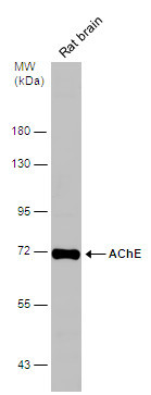
- Submitted by
- GeneTex (provider)
- Main image
- Experimental details
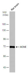
- Rat tissue extract (50 ?g) was separated by 7.5% SDS-PAGE, and the membrane was blotted with AChE antibody (GTX101648) diluted at 1:500.
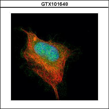
Supportive validation
- Submitted by
- GeneTex (provider)
- Main image
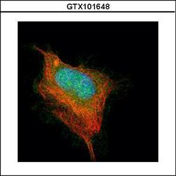
- Experimental details
- Confocal immunofluorescence analysis (Olympus FV10i) of paraformaldehyde-fixed HeLa, using AChE(GTX101648) antibody (Green) at 1:500 dilution. Alpha-tubulin filaments were labeled with GTX11304 (Red) at 1:2500.
Supportive validation
- Submitted by
- GeneTex (provider)
- Main image
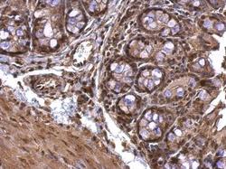
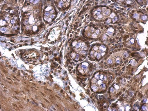
- Experimental details
- AChE antibody detects AChE protein at nucleus on mouse colon by immunohistochemical analysis. Sample: Paraffin-embedded mouse colon. AChE antibody (GTX101648) dilution: 1:500.
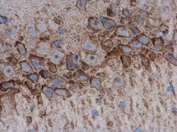
- Submitted by
- GeneTex (provider)
- Main image
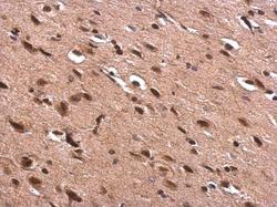
- Experimental details
- AChE antibody detects AChE protein at nucleus on rat fore brain by immunohistochemical analysis. Sample: Paraffin-embedded rat fore brain. AChE antibody (GTX101648) dilution: 1:500.
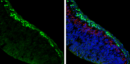
- Submitted by
- GeneTex (provider)
- Main image
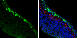
- Experimental details
- AChE antibody detects AChE protein expression by immunohistochemical analysis.Sample: Frozen sectioned E13.5 Rat brain. Green: AChE protein stained by AChE antibody (GTX101648) diluted at 1:250.Red: beta Tubulin 3/ TUJ1, a mature neuron marker, stained by beta Tubulin 3/ TUJ1 antibody [GT11710] (GTX631836) diluted at 1:500.Blue: Fluoroshield with DAPI (GTX30920).
- Submitted by
- GeneTex (provider)
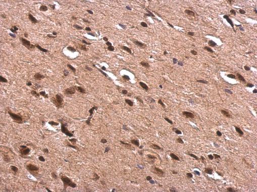
- Main image
- Experimental details
- AChE antibody detects AChE protein at cytoplasm and nucleus in rat brain by immunohistochemical analysis. Sample: Paraffin-embedded rat brain. AChE antibody (GTX101648) diluted at 1:500.
