Explore
 Validate
 Learn
Learn Western blot
 Immunocytochemistry
Antibody data
- Antibody Data
- Antigen structure
- References [0]
- Comments [0]
- Validations
- Western blot [4]
- Immunohistochemistry [4]
Submit
Validation data
Reference
Comment
Report error
- Product number
- NBP1-31329 - Provider product page

- Provider
- Novus Biologicals
- Proper citation
- Novus Cat#NBP1-31329, RRID:AB_2223463
- Product name
- Rabbit Polyclonal Acetylcholinesterase/ACHE Antibody
- Antibody type
- Polyclonal
- Description
- Immunogen affinity purified.
- Reactivity
- Human, Mouse, Rat
- Host
- Rabbit
- Isotype
- IgG
- Vial size
- 100 ul
- Storage
- Aliquot and store at -20C or -80C. Avoid freeze-thaw cycles.
No comments: Submit comment
Supportive validation
- Submitted by
- Novus Biologicals (provider)
- Main image
- Experimental details
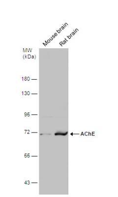
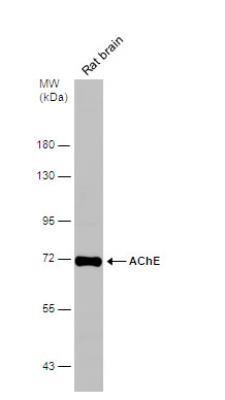
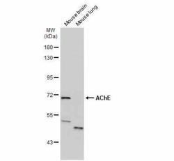
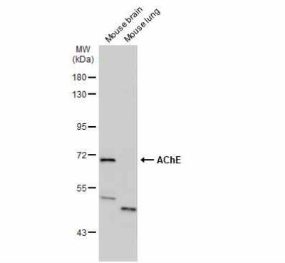
- Western Blot: Acetylcholinesterase/ACHE Antibody [NBP1-31329] - Various tissue extracts (50 ug) were separated by 7.5% SDS-PAGE, and the membrane was blotted with AChE antibody diluted at 1:500. The HRP-conjugated anti-rabbit IgG antibody (NBP2-19301) was used to detect the primary antibody.
- Submitted by
- Novus Biologicals (provider)
- Main image

- Experimental details
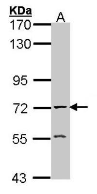
- Western Blot: Acetylcholinesterase/ACHE Antibody [NBP1-31329] - Sample (30 ug of whole cell lysate) A: Raji 7. 5% SDS PAGE; antibody diluted at 1:1000.
- Submitted by
- Novus Biologicals (provider)
- Main image
- Experimental details
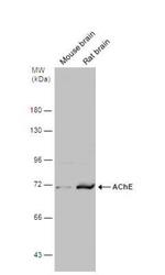
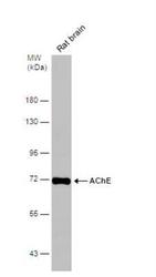
- Western Blot: Acetylcholinesterase/ACHE Antibody [NBP1-31329] - Rat tissue extract (50 ug) was separated by 7.5% SDS-PAGE, and the membrane was blotted with AChE antibody diluted at 1:500.
- Submitted by
- Novus Biologicals (provider)
- Main image
- Experimental details
- Western Blot: Acetylcholinesterase/ACHE Antibody [NBP1-31329] - Various tissue extracts (50 ug) were separated by 7.5% SDS-PAGE, and the membrane was blotted with AChE antibody diluted at 1:500. The HRP-conjugated anti-rabbit IgG antibody (NBP2-19301) was used to detect the primary antibody.
Supportive validation
- Submitted by
- Novus Biologicals (provider)
- Main image
- Experimental details
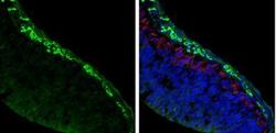
- Immunohistochemistry-Frozen: Acetylcholinesterase/ACHE Antibody [NBP1-31329] - Frozen sectioned E13.5 Rat brain. Green: AChE protein stained by AChE antibody diluted at 1:250. Red: beta Tubulin 3/ TUJ1, a mature neuron marker, stained by beta Tubulin 3/ TUJ1 antibody [11710] (NBP2-43559) diluted at 1:500. Blue: Fluoroshield with DAPI.

- Submitted by
- Novus Biologicals (provider)
- Main image
- Experimental details
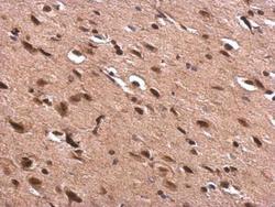
- Immunohistochemistry-Paraffin: Acetylcholinesterase/ACHE Antibody [NBP1-31329] - Rat fore brain. AChE antibody dilution: 1:500. Antigen Retrieval: Trilogy™ (EDTA based, pH 8.0) buffer, 15min.
- Submitted by
- Novus Biologicals (provider)
- Main image
- Experimental details
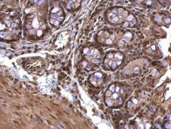
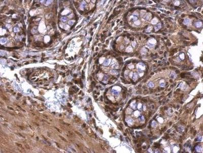
- Immunohistochemistry-Paraffin: Acetylcholinesterase/ACHE Antibody [NBP1-31329] - Mouse colon. AChE antibody dilution: 1:500. Antigen Retrieval: Trilogy™ (EDTA based, pH 8.0) buffer, 15min.
- Submitted by
- Novus Biologicals (provider)
- Main image
- Experimental details
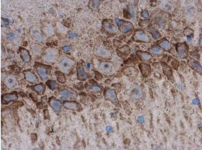
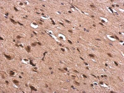
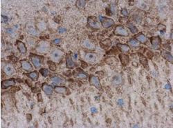
- Immunohistochemistry-Paraffin: Acetylcholinesterase/ACHE Antibody [NBP1-31329] - Rat brain. AChE antibody diluted at 1:500. Antigen Retrieval: Citrate buffer, pH 6.0, 15 min.