Explore
 Validate
 Learn
Learn Western blot
Antibody data
- Antibody Data
- Antigen structure
- References [0]
- Comments [0]
- Validations
- Western blot [1]
- Immunohistochemistry [4]
Submit
Validation data
Reference
Comment
Report error
- Product number
- NBP1-81307 - Provider product page

- Provider
- Novus Biologicals
- Proper citation
- Novus Cat#NBP1-81307, RRID:AB_11003838
- Product name
- Rabbit Polyclonal PLOD3 Antibody
- Antibody type
- Polyclonal
- Description
- Immunogen affinity purified. Specificity of human PLOD3 antibody verified on a Protein Array containing target protein plus 383 other non-specific proteins.
- Reactivity
- Human
- Host
- Rabbit
- Isotype
- IgG
- Vial size
- 0.1 ml
- Storage
- Store at 4C short term. Aliquot and store at -20C long term. Avoid freeze-thaw cycles.
No comments: Submit comment
Supportive validation
- Submitted by
- Novus Biologicals (provider)
- Main image
- Experimental details
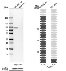
- Western Blot: PLOD3 Antibody [NBP1-81307] - Analysis in human cell lines SK-MEL-30 and HEK293 using Anti-PLOD3 antibody. Corresponding PLOD3 RNA-seq data are presented for the same cell lines. Loading control: Anti-PFN1.

Supportive validation
- Submitted by
- Novus Biologicals (provider)
- Main image
- Experimental details
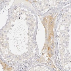
- Immunohistochemistry-Paraffin: PLOD3 Antibody [NBP1-81307] - Staining of human testis shows weak to moderate cytoplasmic positivity in Leydig cells.

- Submitted by
- Novus Biologicals (provider)
- Main image

- Experimental details
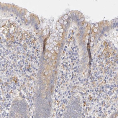
- Immunohistochemistry-Paraffin: PLOD3 Antibody [NBP1-81307] - Staining of human appendix shows weak to moderate cytoplasmic positivity in glandular cells.
- Submitted by
- Novus Biologicals (provider)
- Main image
- Experimental details
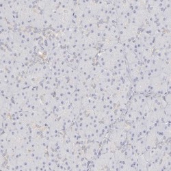
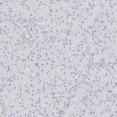
- Immunohistochemistry-Paraffin: PLOD3 Antibody [NBP1-81307] - Staining of human pancreas shows no positivity in exocrine glandular cells.
- Submitted by
- Novus Biologicals (provider)
- Main image
- Experimental details
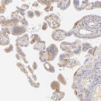
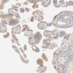
- Immunohistochemistry-Paraffin: PLOD3 Antibody [NBP1-81307] - Staining of human placenta shows weak to moderate cytoplasmic positivity in trophoblastic cells.