 Explore
 Validate
 Learn
Learn Western blot
 ELISA
ELISAAntibody data
- Antibody Data
- Antigen structure
- References [5]
- Comments [0]
- Validations
- Western blot [1]
- Immunocytochemistry [2]
- Other assay [8]
Submit
Validation data
Reference
Comment
Report error
- Product number
- PA1-932 - Provider product page

- Provider
- Invitrogen Antibodies
- Product name
- S100A1 Polyclonal Antibody
- Antibody type
- Polyclonal
- Antigen
- Purifed from natural sources
- Description
- PA1-932 detects S100-alpha protein in bovine, human, mouse, porcine, and rat samples. This antibody has shown no cross-reactivity with the S100-beta protein. PA1-932 has successfully been used in Western blot, ELISA, immunofluorescence, immunocytochemistry, immunoprecipitation and immunohistochemical procedures. By Western blot, this antibody detects a 10 kDa protein representing S100-alpha from human muscle. Immunohistochemical staining on paraffin sections demonstrates that S100-alpha is localized in serous cells, myoepithelial cells and epithelial cells of intercalated ducts in the parotid gland after staining with PA1-932. S100 protein is relatively small and, therefore, it is recommended that the electrophoresis be performed using tricine-SDS-PAGE gels and transferred to a nylon membrane. The PA1-932 immunizing protein corresponds to purified S100-alpha protein from human pectoral muscle cells. Reconstitute in 100 µL PBS to create a stock of 1 mg/mL.
- Reactivity
- Human, Mouse, Rat, Bovine, Porcine
- Host
- Rabbit
- Isotype
- IgG
- Vial size
- 100 μg
- Concentration
- 1 mg/mL
- Storage
- -20°C, Avoid Freeze/Thaw Cycles
Submitted references Evidence of a Myenteric Plexus Barrier and Its Macrophage-Dependent Degradation During Murine Colitis: Implications in Enteric Neuroinflammation.
S100B + A1 CELISA: A Novel Potency Assay and Screening Tool for Redifferentiation Stimuli of Human Articular Chondrocytes.
Progressive changes in detrusor function and micturition patterns with chronic bladder ischemia.
S100A1 and S100B expression patterns identify differentiation status of human articular chondrocytes.
Comparative proteomics profiling of a phospholamban mutant mouse model of dilated cardiomyopathy reveals progressive intracellular stress responses.
Dora D, Ferenczi S, Stavely R, Toth VE, Varga ZV, Kovacs T, Bodi I, Hotta R, Kovacs KJ, Goldstein AM, Nagy N
Cellular and molecular gastroenterology and hepatology 2021;12(5):1617-1641
Cellular and molecular gastroenterology and hepatology 2021;12(5):1617-1641
S100B + A1 CELISA: A Novel Potency Assay and Screening Tool for Redifferentiation Stimuli of Human Articular Chondrocytes.
Diaz-Romero J, Kürsener S, Kohl S, Nesic D
Journal of cellular physiology 2017 Jun;232(6):1559-1570
Journal of cellular physiology 2017 Jun;232(6):1559-1570
Progressive changes in detrusor function and micturition patterns with chronic bladder ischemia.
Zhao Z, Azad R, Yang JH, Siroky MB, Azadzoi KM
Investigative and clinical urology 2016 Jul;57(4):249-59
Investigative and clinical urology 2016 Jul;57(4):249-59
S100A1 and S100B expression patterns identify differentiation status of human articular chondrocytes.
Diaz-Romero J, Quintin A, Schoenholzer E, Pauli C, Despont A, Zumstein MA, Kohl S, Nesic D
Journal of cellular physiology 2014 Aug;229(8):1106-17
Journal of cellular physiology 2014 Aug;229(8):1106-17
Comparative proteomics profiling of a phospholamban mutant mouse model of dilated cardiomyopathy reveals progressive intracellular stress responses.
Gramolini AO, Kislinger T, Alikhani-Koopaei R, Fong V, Thompson NJ, Isserlin R, Sharma P, Oudit GY, Trivieri MG, Fagan A, Kannan A, Higgins DG, Huedig H, Hess G, Arab S, Seidman JG, Seidman CE, Frey B, Perry M, Backx PH, Liu PP, MacLennan DH, Emili A
Molecular & cellular proteomics : MCP 2008 Mar;7(3):519-33
Molecular & cellular proteomics : MCP 2008 Mar;7(3):519-33
No comments: Submit comment
Supportive validation
- Submitted by
- Invitrogen Antibodies (provider)
- Main image
- Experimental details
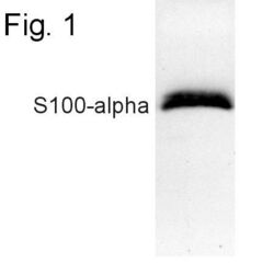
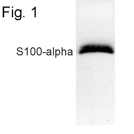
- Western blot of S100-alpha from human muscle extract using Product # PA1-932.
Supportive validation
- Submitted by
- Invitrogen Antibodies (provider)
- Main image
- Experimental details
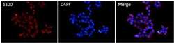
- Immunofluorescent analysis of S100 alpha (red) in HEK293T cells. Cells fixed with 4% formaldehyde were permeabilized and blocked with 1X PBS containing 5% BSA and 0.3% Triton X-100 for 1 hour at room temperature. Cells were probed with a S100 alpha polyclonal antibody (Product # PA1-932) at a dilution of 1:100 overnight at 4°C in 1X PBS containing 1% BSA and 0.3% Triton X-100, washed with 1X PBS, and incubated with a fluorophore-conjugated goat anti-rabbit IgG secondary antibody at a dilution of 1:200 for 1 hour at room temperature. Nuclei (blue) were stained with DAPI. Images were taken on a Leica DM1000 microscope at 40X magnification. Data courtesy of the Innovators Program.

- Submitted by
- Invitrogen Antibodies (provider)
- Main image

- Experimental details
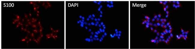
- Immunofluorescent analysis of S100 alpha (red) in HEK293T cells. Cells fixed with 4% formaldehyde were permeabilized and blocked with 1X PBS containing 5% BSA and 0.3% Triton X-100 for 1 hour at room temperature. Cells were probed with a S100 alpha polyclonal antibody (Product # PA1-932) at a dilution of 1:100 overnight at 4°C in 1X PBS containing 1% BSA and 0.3% Triton X-100, washed with 1X PBS, and incubated with a fluorophore-conjugated goat anti-rabbit IgG secondary antibody at a dilution of 1:200 for 1 hour at room temperature. Nuclei (blue) were stained with DAPI. Images were taken on a Leica DM1000 microscope at 40X magnification. Data courtesy of the Innovators Program.
Supportive validation
- Submitted by
- Invitrogen Antibodies (provider)
- Main image
- Experimental details
- NULL
- Submitted by
- Invitrogen Antibodies (provider)
- Main image
- Experimental details
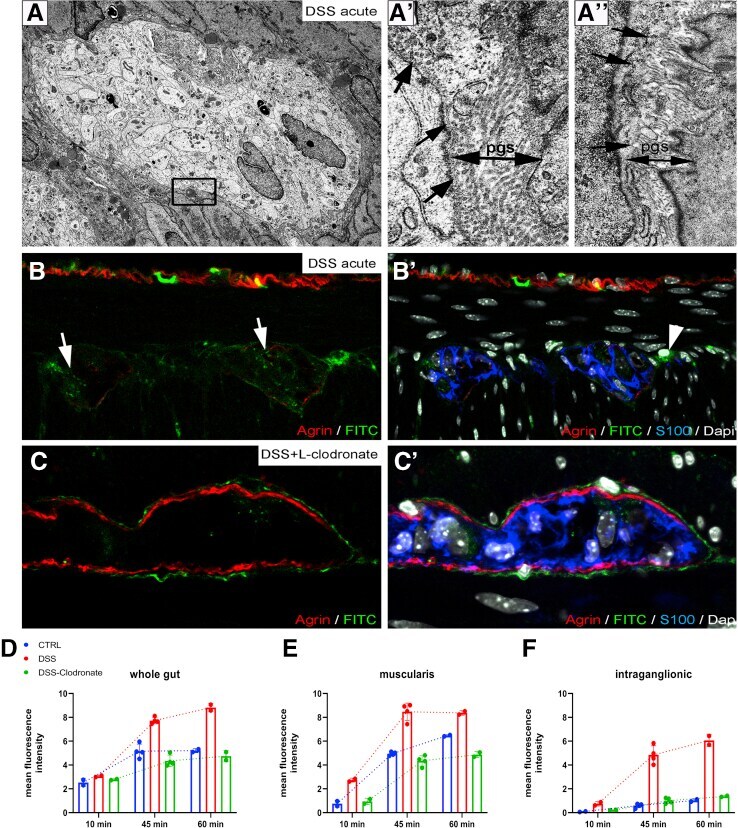
- Figure 12 DSS colitis is associated with macrophage-dependent structural and functional disruption of the BMB. Electron microscopy of DSS-treated colon ( A ) reveals partial disruption ( A', arrows ) or complete absence ( A"", arrows ) of periganglionic basement membrane. PGS is dense and closely packed with collagen fibers ( A'-A"", double arrows ). 60 minutes after administration of FITC-dextran to DSS-treated animals leads to FITC accumulation in the enteric ganglia ( B-B' ) and MyMs ( B', arrowhead ). Clodronate treatment prevents this, leaving FITC-dextran particles accumulating in the PGS surrounding the ganglia ( C-C' ). D , E , and F show the change in mean fluorescent intensity 10, 45, and 60 minutes after FITC-dextran injections in whole gut ( D ), muscularis ( E ), and enteric ganglia ( F ).
- Submitted by
- Invitrogen Antibodies (provider)
- Main image
- Experimental details
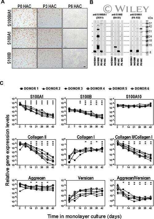
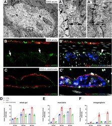
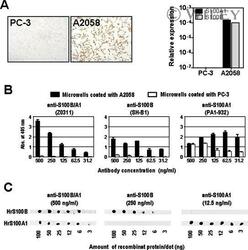
- Characterization of anti-S100A1 and anti-S100B antibodies by cell-based ELISA and dot blot. A: Immunocytochemistry with the anti-S100B/A1 antibody (Z0311; left) and relative expression levels (normalized to 18S) of S100A1 and S100B genes measured by qRT-PCR (right) of the human cell lines A2058 (expressing S100B and S100A1) and PC-3 (negative for both) used for the cell-based ELISA. B: Titration of rabbit polyclonal Z0311 (S100B- and S100A1-specific), mouse monoclonal SH-B1 (S100B-specific), and rabbit polyclonal PA1-932 (S100A1-specific) antibodies by cell-based ELISA using microplates coated with A2058 and PC-3. C: Evaluation of the specificity of the anti-S100 antibodies by dot blot using different concentrations of human recombinant S100A1 (HrS100A1) and S100B (HrS100B).

- Submitted by
- Invitrogen Antibodies (provider)
- Main image
- Experimental details
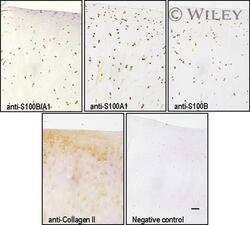
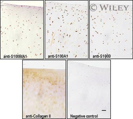
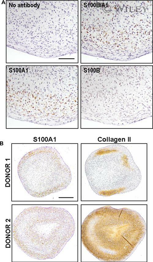
- Expression of S100A1 and S100B in human articular cartilage. Normal human articular cartilage tissue sections were stained with anti-S100B/A1, anti-S100A1, anti-S100B, and collagen type II. A negative control, where the primary antibody was omitted, is shown. Bar = 100 mum.
- Submitted by
- Invitrogen Antibodies (provider)
- Main image
- Experimental details
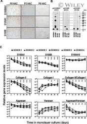
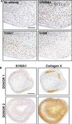
- Re-expression of S100A1 during HAC redifferentiation in pellet culture. P6 HAC incubated in pellet cultures for 3 weeks in serum-free medium with TGF-beta1 and dexamethasone were stained with anti-S100B/A1, anti-S100A1, or anti-S100B antibodies (A) or with anti-S100A1 (lower magnification of A) and anti-collagen type II (B). Representative results from one (A) or two (B) from three different donors are shown. Bar = 100 mum for (A) and 500 mum for (B).
- Submitted by
- Invitrogen Antibodies (provider)
- Main image
- Experimental details
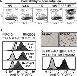
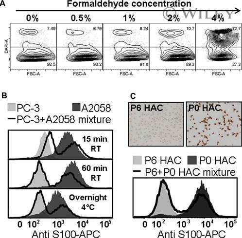
- Development of an intracellular flow cytometry assay for S100 proteins. A: Biparametric histograms of forward scatter (FSC) versus DAPI (nuclear staining) of PC-3 cells showing the effect of formaldehyde concentration used for cell fixation on cell permeability; cells in the upper quadrant (DAPI+) represent permeabilized cells. B: Effects of fixation time and temperature on S100 intracellular stain; PC-3, A2058, and a mixture thereof were fixed with 2% formaldehyde for either 15 min at RT, 60 min at RT, or overnight at 4degC, permeabilized with saponin, and stained with anti-S100-APC. C: Suitability of the method to distinguish differentiated and dedifferentiated HAC based on S100 stain. HAC cultured in monolayer for 2 (P0), 42 (P6) days, and a mixture thereof were fixed with 2% formaldehyde for 60 min at RT, permeabilized with saponin and stained with anti-S100-APC.
- Submitted by
- Invitrogen Antibodies (provider)
- Main image

- Experimental details
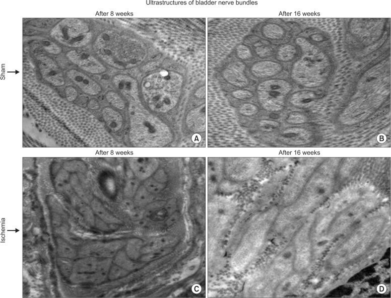
- Fig. 5 Transmission electron microscopy of nerve bundles after 8- (C) and 16-week (D) bladder ischemia are shown versus corresponding sham controls (A and B, respectively). Disrupted axonal ultrastructures after 8-week bladder ischemia and swollen degenerating axons with considerable connective tissue proliferation around the nerve bundles are shown after 16-week bladder ischemia (x9,300).
- Submitted by
- Invitrogen Antibodies (provider)
- Main image
- Experimental details
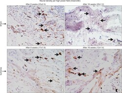
- Fig. 6 Immunoreactive nerve fibers in the ischemic (C and D) and sham control (A and B) bladder tissues are shown at x400 magnifications. Progressive decrease in neural density was evident with increasing duration of ischemia. The number of immunopositive nerves decreased after 8-week bladder ischemia but did not reach significance. After 16-week bladder ischemia, immunoreactive nerve fibers density significantly decreased versus sham control. Arrows point to immunopositive nerve fibers. SE, standard error.
