Explore
 Validate
 Learn
Learn Western blot
 Immunohistochemistry
Antibody data
- Antibody Data
- Antigen structure
- References [0]
- Comments [0]
- Validations
- Immunohistochemistry [23]
Submit
Validation data
Reference
Comment
Report error
- Product number
- LS-C95186 - Provider product page

- Provider
- LSBio
- Product name
- TPC2 / TPCN2 Antibody (aa700-752) LS-C95186
- Antibody type
- Polyclonal
- Description
- Purified
- Reactivity
- Human
- Host
- Rabbit
- Isotype
- IgG
- Storage
- Maintain lyophilized and reconstituted antibodies at -20°C for long term storage and at 2°C to 8°C for a shorter term. When reconstituting, glycerol (1:1) may be added for an additional stability. Avoid freeze/thaw cycles.
No comments: Submit comment
Enhanced validation
- Submitted by
- LSBio (provider)
- Enhanced method
- Genetic validation
- Main image

- Experimental details
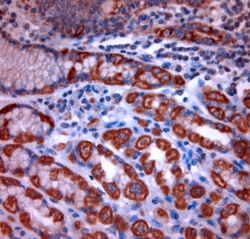
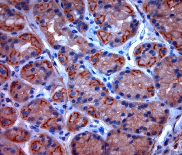
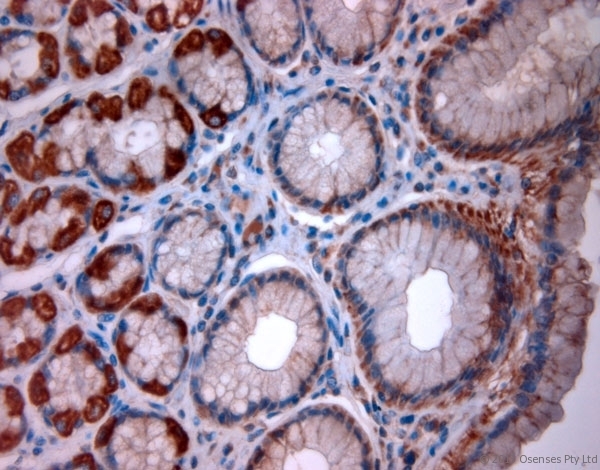
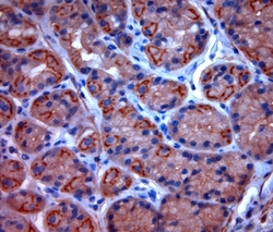
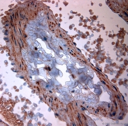
- Rabbit antibody to TPC2 (700-752). IHC-P on paraffin sections of human stomach. HIER: Tris-EDTA, pH 9 for 20 min using Thermo PT Module. Blocking: 0.2% LFDM in TBST filtered through a 0.2 micron filter. Detection was done using Novolink HRP polymer from Leica following manufacturers instructions. Primary antibody: dilution 10 ug/ml, incubated 30 min at RT using Autostainer. Sections were counterstained with Harris Hematoxylin.

- Submitted by
- LSBio (provider)
- Enhanced method
- Genetic validation
- Main image
- Experimental details
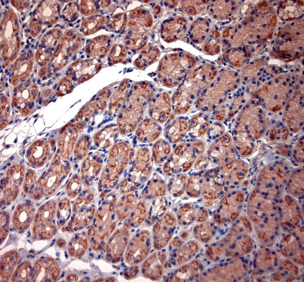
- Rabbit antibody to TPC2 (700-752). IHC-P on paraffin sections of human stomach. HIER: Tris-EDTA, pH 9 for 20 min using Thermo PT Module. Blocking: 0.2% LFDM in TBST filtered through a 0.2 micron filter. Detection was done using Novolink HRP polymer from Leica following manufacturers instructions. Primary antibody: dilution 10 ug/ml, incubated 30 min at RT using Autostainer. Sections were counterstained with Harris Hematoxylin.

- Submitted by
- LSBio (provider)
- Enhanced method
- Genetic validation
- Main image

- Experimental details
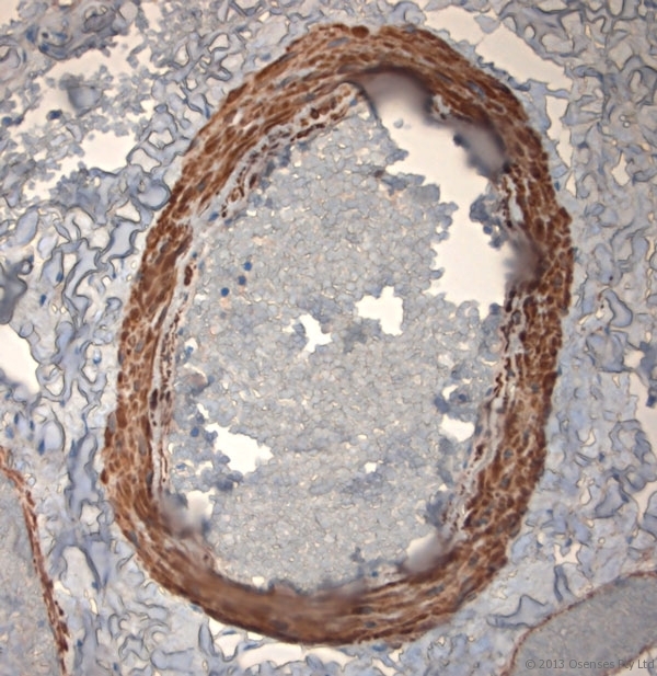
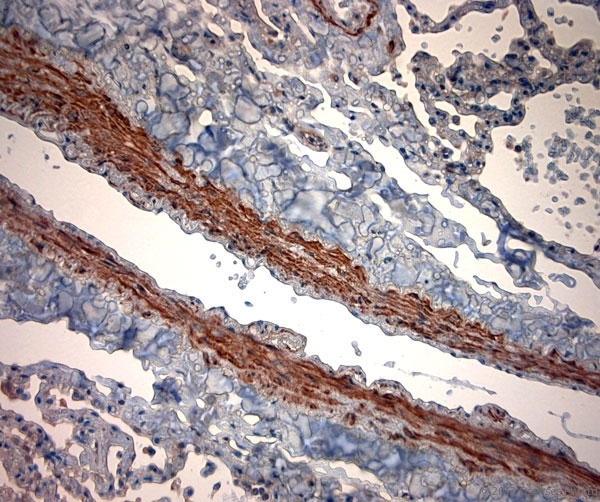
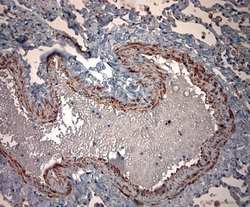
- Rabbit antibody to TPC2 (700-752). IHC-P on paraffin sections of human lung. HIER: Tris-EDTA, pH 9 for 20 min using Thermo PT Module. Blocking: 0.2% LFDM in TBST filtered through a 0.2 micron filter. Detection was done using Novolink HRP polymer from Leica following manufacturers instructions. Primary antibody: dilution 10 ug/ml, incubated 30 min at RT using Autostainer. Sections were counterstained with Harris Hematoxylin.
- Submitted by
- LSBio (provider)
- Enhanced method
- Genetic validation
- Main image

- Experimental details
- Rabbit antibody to TPC2 (700-752). IHC-P on paraffin sections of human stomach. HIER: Tris-EDTA, pH 9 for 20 min using Thermo PT Module. Blocking: 0.2% LFDM in TBST filtered through a 0.2 micron filter. Detection was done using Novolink HRP polymer from Leica following manufacturers instructions. Primary antibody: dilution 10 ug/ml, incubated 30 min at RT using Autostainer. Sections were counterstained with Harris Hematoxylin.

- Submitted by
- LSBio (provider)
- Enhanced method
- Genetic validation
- Main image

- Experimental details
- Rabbit antibody to TPC2 (700-752). IHC-P on paraffin sections of human lung. HIER: Tris-EDTA, pH 9 for 20 min using Thermo PT Module. Blocking: 0.2% LFDM in TBST filtered through a 0.2 micron filter. Detection was done using Novolink HRP polymer from Leica following manufacturers instructions. Primary antibody: dilution 10 ug/ml, incubated 30 min at RT using Autostainer. Sections were counterstained with Harris Hematoxylin.

- Submitted by
- LSBio (provider)
- Enhanced method
- Genetic validation
- Main image
- Experimental details
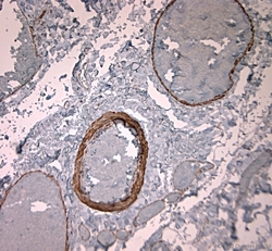
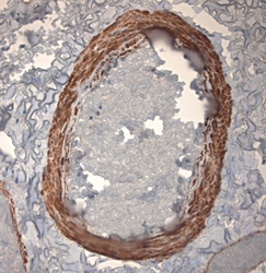
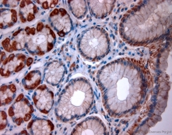
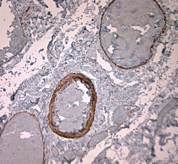
- Rabbit antibody to TPC2 (700-752). IHC-P on paraffin sections of human gallbladder. HIER: Tris-EDTA, pH 9 for 20 min using Thermo PT Module. Blocking: 0.2% LFDM in TBST filtered through a 0.2 micron filter. Detection was done using Novolink HRP polymer from Leica following manufacturers instructions. Primary antibody: dilution 10 ug/ml, incubated 30 min at RT using Autostainer. Sections were counterstained with Harris Hematoxylin.

- Submitted by
- LSBio (provider)
- Enhanced method
- Genetic validation
- Main image
- Experimental details
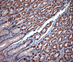
- Rabbit antibody to TPC2 (700-752). IHC-P on paraffin sections of human gallbladder. HIER: Tris-EDTA, pH 9 for 20 min using Thermo PT Module. Blocking: 0.2% LFDM in TBST filtered through a 0.2 micron filter. Detection was done using Novolink HRP polymer from Leica following manufacturers instructions. Primary antibody: dilution 10 ug/ml, incubated 30 min at RT using Autostainer. Sections were counterstained with Harris Hematoxylin.

- Submitted by
- LSBio (provider)
- Main image
- Experimental details
- Rabbit antibody to TPC2 (700-752). IHC-P on paraffin sections of human stomach. HIER: Tris-EDTA, pH 9 for 20 min using Thermo PT Module. Blocking: 0.2% LFDM in TBST filtered through a 0.2 micron filter. Detection was done using Novolink HRP polymer from Leica following manufacturers instructions. Primary antibody: dilution 10 ug/ml, incubated 30 min at RT using Autostainer. Sections were counterstained with Harris Hematoxylin.

- Submitted by
- LSBio (provider)
- Main image

- Experimental details
- Rabbit antibody to TPC2 (700-752). IHC-P on paraffin sections of human stomach. HIER: Tris-EDTA, pH 9 for 20 min using Thermo PT Module. Blocking: 0.2% LFDM in TBST filtered through a 0.2 micron filter. Detection was done using Novolink HRP polymer from Leica following manufacturers instructions. Primary antibody: dilution 10 ug/ml, incubated 30 min at RT using Autostainer. Sections were counterstained with Harris Hematoxylin.
- Submitted by
- LSBio (provider)
- Main image
- Experimental details
- Rabbit antibody to TPC2 (700-752). IHC-P on paraffin sections of human stomach. HIER: Tris-EDTA, pH 9 for 20 min using Thermo PT Module. Blocking: 0.2% LFDM in TBST filtered through a 0.2 micron filter. Detection was done using Novolink HRP polymer from Leica following manufacturers instructions. Primary antibody: dilution 10 ug/ml, incubated 30 min at RT using Autostainer. Sections were counterstained with Harris Hematoxylin.

- Submitted by
- LSBio (provider)
- Main image

- Experimental details
- Rabbit antibody to TPC2 (700-752). IHC-P on paraffin sections of human lung. HIER: Tris-EDTA, pH 9 for 20 min using Thermo PT Module. Blocking: 0.2% LFDM in TBST filtered through a 0.2 micron filter. Detection was done using Novolink HRP polymer from Leica following manufacturers instructions. Primary antibody: dilution 10 ug/ml, incubated 30 min at RT using Autostainer. Sections were counterstained with Harris Hematoxylin.

- Submitted by
- LSBio (provider)
- Main image

- Experimental details
- Rabbit antibody to TPC2 (700-752). IHC-P on paraffin sections of human gallbladder. HIER: Tris-EDTA, pH 9 for 20 min using Thermo PT Module. Blocking: 0.2% LFDM in TBST filtered through a 0.2 micron filter. Detection was done using Novolink HRP polymer from Leica following manufacturers instructions. Primary antibody: dilution 10 ug/ml, incubated 30 min at RT using Autostainer. Sections were counterstained with Harris Hematoxylin.