 Explore
 Validate
 Learn
Learn Western blot
Antibody data
- Antibody Data
- Antigen structure
- References [0]
- Comments [0]
- Validations
- Western blot [1]
- Immunocytochemistry [1]
- Immunohistochemistry [2]
Submit
Validation data
Reference
Comment
Report error
- Product number
- ACC-072-25UL - Provider product page

- Provider
- Invitrogen Antibodies
- Product name
- TPCN2 Polyclonal Antibody
- Antibody type
- Polyclonal
- Antigen
- Other
- Reactivity
- Human, Mouse, Rat
- Host
- Rabbit
- Isotype
- IgG
- Vial size
- 25 µL
- Concentration
- 0.8 mg/mL
- Storage
- -20° C, Avoid Freeze/Thaw Cycles
No comments: Submit comment
Supportive validation
- Submitted by
- Invitrogen Antibodies (provider)
- Main image
- Experimental details
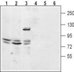
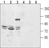
- Western blot analysis of mouse kidney lysate (lanes 1 and 4), rat lung membrane (lanes 2 and 5) and human embryonic Kidney 293 cell lysate (lanes 3 and 6): - 1-3. Anti-TPCN2 Antibody (#ACC-072), (1:200).4-6. Anti-TPCN2 Antibody , preincubated with TPCN2 Blocking Peptide (#BLP-CC072).
Supportive validation
- Submitted by
- Invitrogen Antibodies (provider)
- Main image
- Experimental details
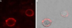
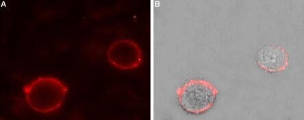
- Expression of Two pore calcium channel protein 2 in rat PC12 cells - Cell surface detection of Two pore calcium channel protein 2 in intact living rat pheochromocytoma (PC12) cells. A. Extracellular staining of cells using Anti-TPCN2 Antibody , (#ACC-072), (1:50), (red). B. Merge of A with the live view of the cell.
Supportive validation
- Submitted by
- Invitrogen Antibodies (provider)
- Main image

- Experimental details
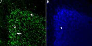
- Expression of Two pore calcium channel protein 2 in rat cerebellum - Immunohistochemical staining of Two pore calcium channel protein 2 in rat cerebellum using Anti-TPCN2 Antibody (#ACC-072).A. TPCN2 positive cells (green)appear in the granule cell layer (G) of the cerebellum (arrows). B. The extent of the granule layer is demonstrated with the counterstain DAPI (blue).
- Submitted by
- Invitrogen Antibodies (provider)
- Main image

- Experimental details
- Expression of Two pore calcium channel protein 2 in rat cerebellum - Immunohistochemical staining of Two pore calcium channel protein 2 in rat cerebellum using Anti-TPCN2 Antibody (#ACC-072).A. TPCN2 positive cells (green)appear in the granule cell layer (G) of the cerebellum (arrows). B. The extent of the granule layer is demonstrated with the counterstain DAPI (blue).
