Explore
 Validate
 Learn
Learn Western blot
 ELISA
ELISAAntibody data
- Antibody Data
- Antigen structure
- References [54]
- Comments [0]
- Validations
- Western blot [1]
- Immunohistochemistry [6]
- Other assay [49]
Submit
Validation data
Reference
Comment
Report error
- Product number
- MA1-934 - Provider product page

- Provider
- Invitrogen Antibodies
- Product name
- Calcium Sensing Receptor Monoclonal Antibody (5C10, ADD)
- Antibody type
- Monoclonal
- Antigen
- Synthetic peptide
- Description
- MA1-934 detects human, bovine, mouse, and rat calcium sensing receptor. MA1-934 has been successfully used in Western blot, immunohistochemistry, and ELISA procedures. By Western blot, this antibody detects an ~ 150 and 130 kDa proteins from human HEK-293 cells.This antibody has also been used in immunohistochemical on frozen bovine parathyroid gland and rat thyroid gland. The MA1-934 immunogen is a synthetic peptide corresponding to residues (214) A D D D Y G R P G I E K F RE E A E E R D I (235) of human calcium sensing receptor.
- Reactivity
- Human, Mouse, Rat, Bovine
- Host
- Mouse
- Isotype
- IgG
- Antibody clone number
- 5C10, ADD
- Vial size
- 100 μg
- Concentration
- 1 mg/mL
- Storage
- -20°C, Avoid Freeze/Thaw Cycles
Submitted references Density-Dependent Differentiation of Tonsil-Derived Mesenchymal Stem Cells into Parathyroid-Hormone-Releasing Cells.
Two cases of aldosterone and cortisol producing adenoma with different histopathological features: A case report.
Urothelial Calcium-Sensing Receptor Modulates Micturition Function via Mediating Detrusor Activity and Ameliorates Bladder Hyperactivity in Rats.
Calcium-Sensing Receptors Control CYP27B1-Luciferase Expression: Transcriptional and Posttranscriptional Mechanisms.
Study of the Expression and Function of Calcium-Sensing Receptor in Human Skeletal Muscle.
In Vitro Control of Genes Critical for Parathyroid Embryogenesis by Extracellular Calcium.
Phosphate acts directly on the calcium-sensing receptor to stimulate parathyroid hormone secretion.
L-phenylalanine Increased Gut Hormone Secretion through Calcium-Sensing Receptor in the Porcine Duodenum.
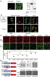
Cinacalcet corrects biased allosteric modulation of CaSR by AHH autoantibody.
Calcium-sensing receptor-mediated L-tryptophan-induced secretion of cholecystokinin and glucose-dependent insulinotropic peptide in swine duodenum.
Peripartum Fluoxetine Reduces Maternal Trabecular Bone After Weaning and Elevates Mammary Gland Serotonin and PTHrP.
Orphan Adhesion GPCR GPR64/ADGRG2 Is Overexpressed in Parathyroid Tumors and Attenuates Calcium-Sensing Receptor-Mediated Signaling.
Common variants in CLDN14 are associated with differential excretion of magnesium over calcium in urine.
Persistent fibroblast growth factor 23 signalling in the parathyroid glands for secondary hyperparathyroidism in mice with chronic kidney disease.
Notch activation of Ca(2+)-sensing receptor mediates hypoxia-induced pulmonary hypertension.
Comparative expression of the extracellular calcium-sensing receptor in the mouse, rat, and human kidney.
pH-sensitive expression of calcium-sensing receptor (CaSR) in type-B intercalated cells of the cortical collecting ducts (CCD) in mouse kidney.
Peripheral serotonin regulates maternal calcium trafficking in mammary epithelial cells during lactation in mice.
PTH-C1: a rat continuous cell line expressing the parathyroid phenotype.
Inhibition of the Ca(2+)-sensing receptor rescues pulmonary hypertension in rats and mice.
Gastric neuropeptide W is regulated by meal-related nutrients.
Molecular mechanisms of calcium-sensing receptor-mediated calcium signaling in the modulation of epithelial ion transport and bicarbonate secretion.
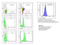
Total calcium-sensing receptor expression in circulating monocytes is increased in rheumatoid arthritis patients with severe coronary artery calcification.
Determination and modulation of total and surface calcium-sensing receptor expression in monocytes in vivo and in vitro.
Calcium-sensing receptor in rat vagal bronchopulmonary sensory neurons regulates the function of the capsaicin receptor TRPV1.
Calcimimetic R-568 and its enantiomer S-568 increase nitric oxide release in human endothelial cells.
Receptors responsive to protein breakdown products in g-cells and d-cells of mouse, swine and human.
PTH-independent regulation of blood calcium concentration by the calcium-sensing receptor.
Calcium-sensing receptor inhibits secretagogue-induced electrolyte secretion by intestine via the enteric nervous system.
The adaptor protein 14-3-3 binds to the calcium-sensing receptor and attenuates receptor-mediated Rho kinase signalling.
Enhanced Ca(2+)-sensing receptor function in idiopathic pulmonary arterial hypertension.
Thermostable direct hemolysin downregulates human colon carcinoma cell proliferation with the involvement of E-cadherin, and β-catenin/Tcf-4 signaling.
Calcium-sensing receptor modulates cell adhesion and migration via integrins.
Regulator of G protein signaling 5 is highly expressed in parathyroid tumors and inhibits signaling by the calcium-sensing receptor.
A novel mutation of the primary protein kinase C phosphorylation site in the calcium-sensing receptor causes autosomal dominant hypocalcemia.
Nutrient sensing receptors in gastric endocrine cells.
The extracellular calcium-sensing receptor is required for cholecystokinin secretion in response to L-phenylalanine in acutely isolated intestinal I cells.
Calcimimetic R-568 effects on activity of R990G polymorphism of calcium-sensing receptor.
Rab1 small GTP-binding protein regulates cell surface trafficking of the human calcium-sensing receptor.
Increased receptor stimulation elicits differential calcium-sensing receptor(T888) dephosphorylation.
Extracellular calcium-sensing receptor mediates human bronchial epithelial wound repair.
Calcium-sensing receptor as a potential modulator of vascular calcification in chronic kidney disease.
Nickel induces intracellular calcium mobilization and pathophysiological responses in human cultured airway epithelial cells.
Development of a technique for introduction of an expressed complementary deoxyribonucleic acid into parathyroid cells by direct injection.
Calcification is associated with loss of functional calcium-sensing receptor in vascular smooth muscle cells.
Identification of a Ca2+-sensing receptor in rat trigeminal ganglia, sensory axons, and tooth dental pulp.
Ca(2+) mobilization through dorsal root ganglion Ca(2+)-sensing receptor stably expressed in HEK293 cells.
Elucidation of the role of peptide linker in calcium-sensing receptor activation process.
Activation of family C G-protein-coupled receptors by the tripeptide glutathione.
Evidence in favor of a calcium-sensing receptor in arterial endothelial cells: studies with calindol and Calhex 231.
Exercise training improves cardiac function-related gene levels through thyroid hormone receptor signaling in aged rats.
Expression, purification, and biochemical characterization of the amino-terminal extracellular domain of the human calcium receptor.
Expression, purification, and biochemical characterization of the amino-terminal extracellular domain of the human calcium receptor.
Monoclonal antibodies against synthetic peptides corresponding to the extracellular domain of the human Ca2+ receptor: characterization and use in studying concanavalin A inhibition.
Kim JY, Park S, Oh SY, Nam YH, Choi YM, Choi Y, Kim HY, Jung SY, Kim HS, Jo I, Jung SC
International journal of molecular sciences 2022 Jan 10;23(2)
International journal of molecular sciences 2022 Jan 10;23(2)
Two cases of aldosterone and cortisol producing adenoma with different histopathological features: A case report.
Gao H, Li L, Tian H
Medicine 2022 Aug 12;101(32):e30008
Medicine 2022 Aug 12;101(32):e30008
Urothelial Calcium-Sensing Receptor Modulates Micturition Function via Mediating Detrusor Activity and Ameliorates Bladder Hyperactivity in Rats.
Wu WY, Lee SP, Chiang BJ, Lin WY, Chien CT
Pharmaceuticals (Basel, Switzerland) 2021 Sep 23;14(10)
Pharmaceuticals (Basel, Switzerland) 2021 Sep 23;14(10)
Calcium-Sensing Receptors Control CYP27B1-Luciferase Expression: Transcriptional and Posttranscriptional Mechanisms.
Huang A, Binmahfouz L, Hancock DP, Anderson PH, Ward DT, Conigrave AD
Journal of the Endocrine Society 2021 Sep 1;5(9):bvab057
Journal of the Endocrine Society 2021 Sep 1;5(9):bvab057
Study of the Expression and Function of Calcium-Sensing Receptor in Human Skeletal Muscle.
Romagnoli C, Sharma P, Zonefrati R, Palmini G, Lucattelli E, Ward DT, Ellinger I, Innocenti M, Brandi ML
International journal of molecular sciences 2021 Jul 6;22(14)
International journal of molecular sciences 2021 Jul 6;22(14)
In Vitro Control of Genes Critical for Parathyroid Embryogenesis by Extracellular Calcium.
Fabbri S, Zonefrati R, Galli G, Gronchi G, Perigli G, Borrelli A, Brandi ML
Journal of the Endocrine Society 2020 Jul 1;4(7):bvaa058
Journal of the Endocrine Society 2020 Jul 1;4(7):bvaa058
Phosphate acts directly on the calcium-sensing receptor to stimulate parathyroid hormone secretion.
Centeno PP, Herberger A, Mun HC, Tu C, Nemeth EF, Chang W, Conigrave AD, Ward DT
Nature communications 2019 Oct 16;10(1):4693
Nature communications 2019 Oct 16;10(1):4693
L-phenylalanine Increased Gut Hormone Secretion through Calcium-Sensing Receptor in the Porcine Duodenum.
Feng J, Kang C, Wang C, Ding L, Zhu W, Hang S
Animals : an open access journal from MDPI 2019 Jul 24;9(8)
Animals : an open access journal from MDPI 2019 Jul 24;9(8)
Cinacalcet corrects biased allosteric modulation of CaSR by AHH autoantibody.
Makita N, Ando T, Sato J, Manaka K, Mitani K, Kikuchi Y, Niwa T, Ootaki M, Takeba Y, Matsumoto N, Kawakami A, Ogawa T, Nangaku M, Iiri T
JCI insight 2019 Apr 18;4(8)
JCI insight 2019 Apr 18;4(8)
Calcium-sensing receptor-mediated L-tryptophan-induced secretion of cholecystokinin and glucose-dependent insulinotropic peptide in swine duodenum.
Zhao X, Xian Y, Wang C, Ding L, Meng X, Zhu W, Hang S
Journal of veterinary science 2018 Mar 31;19(2):179-187
Journal of veterinary science 2018 Mar 31;19(2):179-187
Peripartum Fluoxetine Reduces Maternal Trabecular Bone After Weaning and Elevates Mammary Gland Serotonin and PTHrP.
Weaver SR, Fricke HP, Xie C, Lipinski RJ, Vezina CM, Charles JF, Hernandez LL
Endocrinology 2018 Aug 1;159(8):2850-2862
Endocrinology 2018 Aug 1;159(8):2850-2862
Orphan Adhesion GPCR GPR64/ADGRG2 Is Overexpressed in Parathyroid Tumors and Attenuates Calcium-Sensing Receptor-Mediated Signaling.
Balenga N, Azimzadeh P, Hogue JA, Staats PN, Shi Y, Koh J, Dressman H, Olson JA Jr
Journal of bone and mineral research : the official journal of the American Society for Bone and Mineral Research 2017 Mar;32(3):654-666
Journal of bone and mineral research : the official journal of the American Society for Bone and Mineral Research 2017 Mar;32(3):654-666
Common variants in CLDN14 are associated with differential excretion of magnesium over calcium in urine.
Corre T, Olinger E, Harris SE, Traglia M, Ulivi S, Lenarduzzi S, Belge H, Youhanna S, Tokonami N, Bonny O, Houillier P, Polasek O, Deary IJ, Starr JM, Toniolo D, Gasparini P, Vollenweider P, Hayward C, Bochud M, Devuyst O
Pflugers Archiv : European journal of physiology 2017 Jan;469(1):91-103
Pflugers Archiv : European journal of physiology 2017 Jan;469(1):91-103
Persistent fibroblast growth factor 23 signalling in the parathyroid glands for secondary hyperparathyroidism in mice with chronic kidney disease.
Kawakami K, Takeshita A, Furushima K, Miyajima M, Hatamura I, Kuro-O M, Furuta Y, Sakaguchi K
Scientific reports 2017 Jan 17;7:40534
Scientific reports 2017 Jan 17;7:40534
Notch activation of Ca(2+)-sensing receptor mediates hypoxia-induced pulmonary hypertension.
Guo Q, Xu H, Yang X, Zhao D, Liu S, Sun X, Huang JA
Hypertension research : official journal of the Japanese Society of Hypertension 2017 Feb;40(2):117-129
Hypertension research : official journal of the Japanese Society of Hypertension 2017 Feb;40(2):117-129
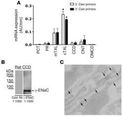
Comparative expression of the extracellular calcium-sensing receptor in the mouse, rat, and human kidney.
Graca JA, Schepelmann M, Brennan SC, Reens J, Chang W, Yan P, Toka H, Riccardi D, Price SA
American journal of physiology. Renal physiology 2016 Mar 15;310(6):F518-33
American journal of physiology. Renal physiology 2016 Mar 15;310(6):F518-33
pH-sensitive expression of calcium-sensing receptor (CaSR) in type-B intercalated cells of the cortical collecting ducts (CCD) in mouse kidney.
Yasuoka Y, Sato Y, Healy JM, Nonoguchi H, Kawahara K
Clinical and experimental nephrology 2015 Oct;19(5):771-82
Clinical and experimental nephrology 2015 Oct;19(5):771-82
Peripheral serotonin regulates maternal calcium trafficking in mammary epithelial cells during lactation in mice.
Laporta J, Keil KP, Vezina CM, Hernandez LL
PloS one 2014;9(10):e110190
PloS one 2014;9(10):e110190
PTH-C1: a rat continuous cell line expressing the parathyroid phenotype.
Fabbri S, Ciuffi S, Nardone V, Gomes AR, Mavilia C, Zonefrati R, Galli G, Luzi E, Tanini A, Brandi ML
Endocrine 2014 Sep;47(1):90-9
Endocrine 2014 Sep;47(1):90-9
Inhibition of the Ca(2+)-sensing receptor rescues pulmonary hypertension in rats and mice.
Guo Q, Huang JA, Yamamura A, Yamamura H, Zimnicka AM, Fernandez R, Yuan JX
Hypertension research : official journal of the Japanese Society of Hypertension 2014 Feb;37(2):116-24
Hypertension research : official journal of the Japanese Society of Hypertension 2014 Feb;37(2):116-24
Gastric neuropeptide W is regulated by meal-related nutrients.
Li H, Feinle-Bisset C, Frisby C, Kentish S, Wittert GA, Page AJ
Peptides 2014 Dec;62:6-14
Peptides 2014 Dec;62:6-14
Molecular mechanisms of calcium-sensing receptor-mediated calcium signaling in the modulation of epithelial ion transport and bicarbonate secretion.
Xie R, Dong X, Wong C, Vallon V, Tang B, Sun J, Yang S, Dong H
The Journal of biological chemistry 2014 Dec 12;289(50):34642-53
The Journal of biological chemistry 2014 Dec 12;289(50):34642-53
Total calcium-sensing receptor expression in circulating monocytes is increased in rheumatoid arthritis patients with severe coronary artery calcification.
Paccou J, Boudot C, Renard C, Liabeuf S, Kamel S, Fardellone P, Massy Z, Brazier M, Mentaverri R
Arthritis research & therapy 2014 Aug 19;16(5):412
Arthritis research & therapy 2014 Aug 19;16(5):412
Determination and modulation of total and surface calcium-sensing receptor expression in monocytes in vivo and in vitro.
Paccou J, Boudot C, Mary A, Kamel S, Drüeke TB, Fardellone P, Massy Z, Brazier M, Mentaverri R
PloS one 2013;8(10):e74800
PloS one 2013;8(10):e74800
Calcium-sensing receptor in rat vagal bronchopulmonary sensory neurons regulates the function of the capsaicin receptor TRPV1.
Gu Q, Vysotskaya ZV, Moss CR 2nd, Kagira MK, Gilbert CA
Experimental physiology 2013 Nov;98(11):1631-42
Experimental physiology 2013 Nov;98(11):1631-42
Calcimimetic R-568 and its enantiomer S-568 increase nitric oxide release in human endothelial cells.
Bonomini M, Giardinelli A, Morabito C, Di Silvestre S, Di Cesare M, Di Pietro N, Sirolli V, Formoso G, Amoroso L, Mariggiò MA, Pandolfi A
PloS one 2012;7(1):e30682
PloS one 2012;7(1):e30682
Receptors responsive to protein breakdown products in g-cells and d-cells of mouse, swine and human.
Haid DC, Jordan-Biegger C, Widmayer P, Breer H
Frontiers in physiology 2012;3:65
Frontiers in physiology 2012;3:65
PTH-independent regulation of blood calcium concentration by the calcium-sensing receptor.
Loupy A, Ramakrishnan SK, Wootla B, Chambrey R, de la Faille R, Bourgeois S, Bruneval P, Mandet C, Christensen EI, Faure H, Cheval L, Laghmani K, Collet C, Eladari D, Dodd RH, Ruat M, Houillier P
The Journal of clinical investigation 2012 Sep;122(9):3355-67
The Journal of clinical investigation 2012 Sep;122(9):3355-67
Calcium-sensing receptor inhibits secretagogue-induced electrolyte secretion by intestine via the enteric nervous system.
Cheng SX
American journal of physiology. Gastrointestinal and liver physiology 2012 Jul;303(1):G60-70
American journal of physiology. Gastrointestinal and liver physiology 2012 Jul;303(1):G60-70
The adaptor protein 14-3-3 binds to the calcium-sensing receptor and attenuates receptor-mediated Rho kinase signalling.
Arulpragasam A, Magno AL, Ingley E, Brown SJ, Conigrave AD, Ratajczak T, Ward BK
The Biochemical journal 2012 Feb 1;441(3):995-1006
The Biochemical journal 2012 Feb 1;441(3):995-1006
Enhanced Ca(2+)-sensing receptor function in idiopathic pulmonary arterial hypertension.
Yamamura A, Guo Q, Yamamura H, Zimnicka AM, Pohl NM, Smith KA, Fernandez RA, Zeifman A, Makino A, Dong H, Yuan JX
Circulation research 2012 Aug 3;111(4):469-81
Circulation research 2012 Aug 3;111(4):469-81
Thermostable direct hemolysin downregulates human colon carcinoma cell proliferation with the involvement of E-cadherin, and β-catenin/Tcf-4 signaling.
Chowdhury P, Pore D, Mahata N, Karmakar P, Pal A, Chakrabarti MK
PloS one 2011;6(5):e20098
PloS one 2011;6(5):e20098
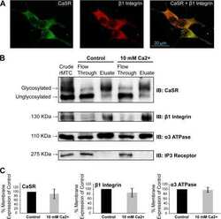
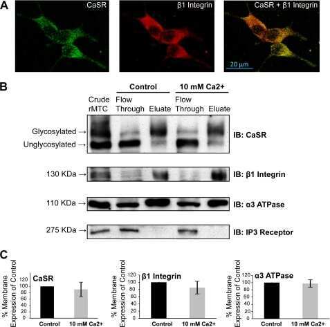
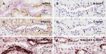
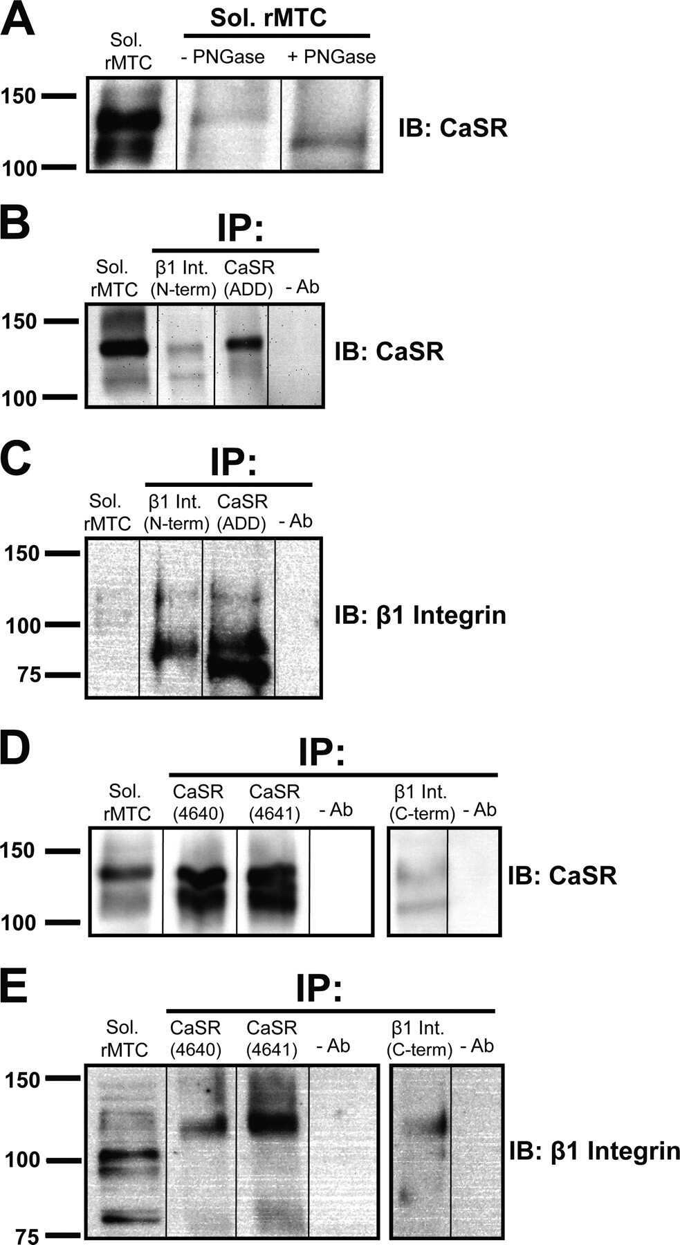
Calcium-sensing receptor modulates cell adhesion and migration via integrins.
Tharmalingam S, Daulat AM, Antflick JE, Ahmed SM, Nemeth EF, Angers S, Conigrave AD, Hampson DR
The Journal of biological chemistry 2011 Nov 25;286(47):40922-33
The Journal of biological chemistry 2011 Nov 25;286(47):40922-33
Regulator of G protein signaling 5 is highly expressed in parathyroid tumors and inhibits signaling by the calcium-sensing receptor.
Koh J, Dar M, Untch BR, Dixit D, Shi Y, Yang Z, Adam MA, Dressman H, Wang X, Gesty-Palmer D, Marks JR, Spurney R, Druey KM, Olson JA Jr
Molecular endocrinology (Baltimore, Md.) 2011 May;25(5):867-76
Molecular endocrinology (Baltimore, Md.) 2011 May;25(5):867-76
A novel mutation of the primary protein kinase C phosphorylation site in the calcium-sensing receptor causes autosomal dominant hypocalcemia.
Lazarus S, Pretorius CJ, Khafagi F, Campion KL, Brennan SC, Conigrave AD, Brown EM, Ward DT
European journal of endocrinology 2011 Mar;164(3):429-35
European journal of endocrinology 2011 Mar;164(3):429-35
Nutrient sensing receptors in gastric endocrine cells.
Haid D, Widmayer P, Breer H
Journal of molecular histology 2011 Aug;42(4):355-64
Journal of molecular histology 2011 Aug;42(4):355-64
The extracellular calcium-sensing receptor is required for cholecystokinin secretion in response to L-phenylalanine in acutely isolated intestinal I cells.
Liou AP, Sei Y, Zhao X, Feng J, Lu X, Thomas C, Pechhold S, Raybould HE, Wank SA
American journal of physiology. Gastrointestinal and liver physiology 2011 Apr;300(4):G538-46
American journal of physiology. Gastrointestinal and liver physiology 2011 Apr;300(4):G538-46
Calcimimetic R-568 effects on activity of R990G polymorphism of calcium-sensing receptor.
Terranegra A, Ferraretto A, Dogliotti E, Scarpellini M, Corbetta S, Barbieri AM, Spada A, Arcidiacono T, Rainone F, Aloia A, Cusi D, Vezzoli G, Soldati L
Journal of molecular endocrinology 2010 Oct;45(4):245-56
Journal of molecular endocrinology 2010 Oct;45(4):245-56
Rab1 small GTP-binding protein regulates cell surface trafficking of the human calcium-sensing receptor.
Zhuang X, Adipietro KA, Datta S, Northup JK, Ray K
Endocrinology 2010 Nov;151(11):5114-23
Endocrinology 2010 Nov;151(11):5114-23
Increased receptor stimulation elicits differential calcium-sensing receptor(T888) dephosphorylation.
McCormick WD, Atkinson-Dell R, Campion KL, Mun HC, Conigrave AD, Ward DT
The Journal of biological chemistry 2010 May 7;285(19):14170-7
The Journal of biological chemistry 2010 May 7;285(19):14170-7
Extracellular calcium-sensing receptor mediates human bronchial epithelial wound repair.
Milara J, Mata M, Serrano A, Peiró T, Morcillo EJ, Cortijo J
Biochemical pharmacology 2010 Jul 15;80(2):236-46
Biochemical pharmacology 2010 Jul 15;80(2):236-46
Calcium-sensing receptor as a potential modulator of vascular calcification in chronic kidney disease.
Caudrillier A, Mentaverri R, Brazier M, Kamel S, Massy ZA
Journal of nephrology 2010 Jan-Feb;23(1):17-22
Journal of nephrology 2010 Jan-Feb;23(1):17-22
Nickel induces intracellular calcium mobilization and pathophysiological responses in human cultured airway epithelial cells.
Cortijo J, Milara J, Mata M, Donet E, Gavara N, Peel SE, Hall IP, Morcillo EJ
Chemico-biological interactions 2010 Jan 5;183(1):25-33
Chemico-biological interactions 2010 Jan 5;183(1):25-33
Development of a technique for introduction of an expressed complementary deoxyribonucleic acid into parathyroid cells by direct injection.
Shiizaki K, Hatamura I, Fukagawa M, Nakazawa E, Saji F, Watanabe Y, Akizawa T, Kusano E
Endocrinology 2010 Aug;151(8):4031-8
Endocrinology 2010 Aug;151(8):4031-8
Calcification is associated with loss of functional calcium-sensing receptor in vascular smooth muscle cells.
Alam MU, Kirton JP, Wilkinson FL, Towers E, Sinha S, Rouhi M, Vizard TN, Sage AP, Martin D, Ward DT, Alexander MY, Riccardi D, Canfield AE
Cardiovascular research 2009 Feb 1;81(2):260-8
Cardiovascular research 2009 Feb 1;81(2):260-8
Identification of a Ca2+-sensing receptor in rat trigeminal ganglia, sensory axons, and tooth dental pulp.
Heyeraas KJ, Haug SR, Bukoski RD, Awumey EM
Calcified tissue international 2008 Jan;82(1):57-65
Calcified tissue international 2008 Jan;82(1):57-65
Ca(2+) mobilization through dorsal root ganglion Ca(2+)-sensing receptor stably expressed in HEK293 cells.
Awumey EM, Howlett AC, Putney JW Jr, Diz DI, Bukoski RD
American journal of physiology. Cell physiology 2007 May;292(5):C1895-905
American journal of physiology. Cell physiology 2007 May;292(5):C1895-905
Elucidation of the role of peptide linker in calcium-sensing receptor activation process.
Ray K, Adipietro KA, Chen C, Northup JK
The Journal of biological chemistry 2007 Feb 23;282(8):5310-7
The Journal of biological chemistry 2007 Feb 23;282(8):5310-7
Activation of family C G-protein-coupled receptors by the tripeptide glutathione.
Wang M, Yao Y, Kuang D, Hampson DR
The Journal of biological chemistry 2006 Mar 31;281(13):8864-70
The Journal of biological chemistry 2006 Mar 31;281(13):8864-70
Evidence in favor of a calcium-sensing receptor in arterial endothelial cells: studies with calindol and Calhex 231.
Weston AH, Absi M, Ward DT, Ohanian J, Dodd RH, Dauban P, Petrel C, Ruat M, Edwards G
Circulation research 2005 Aug 19;97(4):391-8
Circulation research 2005 Aug 19;97(4):391-8
Exercise training improves cardiac function-related gene levels through thyroid hormone receptor signaling in aged rats.
Iemitsu M, Miyauchi T, Maeda S, Tanabe T, Takanashi M, Matsuda M, Yamaguchi I
American journal of physiology. Heart and circulatory physiology 2004 May;286(5):H1696-705
American journal of physiology. Heart and circulatory physiology 2004 May;286(5):H1696-705
Expression, purification, and biochemical characterization of the amino-terminal extracellular domain of the human calcium receptor.
Goldsmith PK, Fan GF, Ray K, Shiloach J, McPhie P, Rogers KV, Spiegel AM
The Journal of biological chemistry 1999 Apr 16;274(16):11303-9
The Journal of biological chemistry 1999 Apr 16;274(16):11303-9
Monoclonal antibodies against synthetic peptides corresponding to the extracellular domain of the human Ca2+ receptor: characterization and use in studying concanavalin A inhibition.
Goldsmith PK, Fan G, Miller JL, Rogers KV, Spiegel AM
Journal of bone and mineral research : the official journal of the American Society for Bone and Mineral Research 1997 Nov;12(11):1780-8
Journal of bone and mineral research : the official journal of the American Society for Bone and Mineral Research 1997 Nov;12(11):1780-8
No comments: Submit comment
Supportive validation
- Submitted by
- Invitrogen Antibodies (provider)
- Main image

- Experimental details
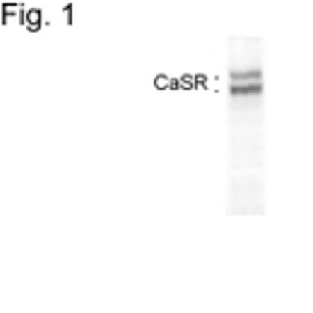
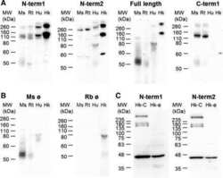
- Western blot analysis of calcium sensing receptor in HEK7-2 cell extract using Calcium Sensing Receptor Monoclonal Antibody (Product # MA1-934).
Supportive validation
- Submitted by
- Invitrogen Antibodies (provider)
- Main image
- Experimental details
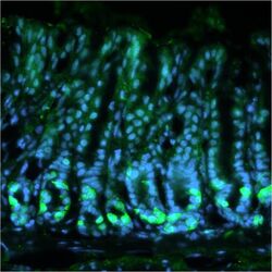
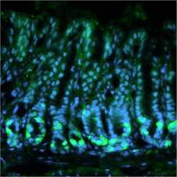
- Immunohistochemical analysis of mouse stomach stained with Calcium Sensing Receptor Monoclonal Antibody (Product # MA1-934). Fresh stomach tissue was fixed in 4% formalin for 1 hour and then incubated overnight at 4°C in 25% sucrose before embedding in tissue freezing medium. Antigen retrieval was carried out on 8 µm cryo-sections by incubating in sodium citrate buffer for 45 minutes at 4°C, immersing in sodium citrate buffer for 10 minutes at 100°C before washing 3 times for 5 minutes each in 1X PBS. Sections were then blocked in blocking buffer (0.3% Triton X-100 in 1X PBS containing 10% normal goat serum) for 30 minutes at RT before staining with Product # MA1-934 (diluted 1:100 in blocking buffer) overnight at 4°C followed by a fluorophore-conjugated goat anti-mouse IgG secondary antibody for 2 hours at RT. Sections were also stained with DAPI nuclear stain (blue). Product # MA1-934 positive cells (green) were found at the base of the antral glands in the mouse stomach. Data courtesy of the Innovators Program.

- Submitted by
- Invitrogen Antibodies (provider)
- Main image

- Experimental details
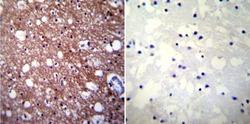
- Immunohistochemistry was performed on normal biopsies of deparaffinized Human brain tissue. To expose target proteins, heat induced antigen retrieval was performed using 10mM sodium citrate (pH6.0) buffer, microwaved for 8-15 minutes. Following antigen retrieval tissues were blocked in 3% BSA-PBS for 30 minutes at room temperature. Tissues were then probed at a dilution of 1:100 with a mouse monoclonal antibody recognizing Calcium Sensing Receptor (Product # MA1-934) or without primary antibody (negative control) overnight at 4°C in a humidified chamber. Tissues were washed extensively with PBST and endogenous peroxidase activity was quenched with a peroxidase suppressor. Detection was performed using a biotin-conjugated secondary antibody and SA-HRP, followed by colorimetric detection using DAB. Tissues were counterstained with hematoxylin and prepped for mounting.

- Submitted by
- Invitrogen Antibodies (provider)
- Main image
- Experimental details
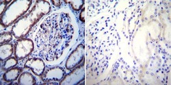
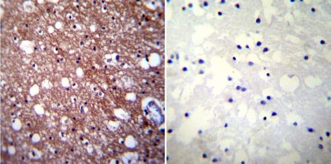
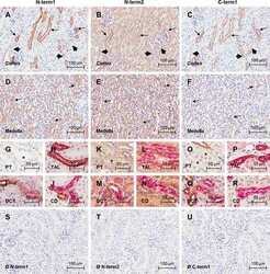
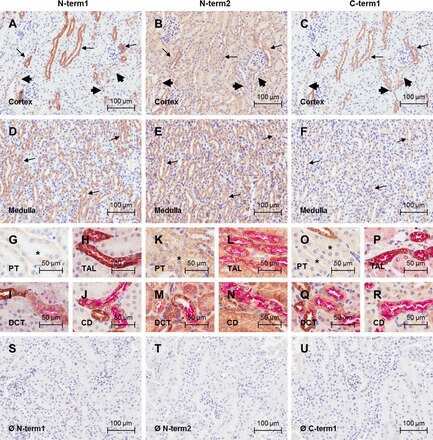
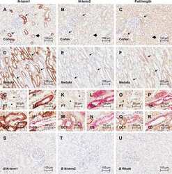
- Immunohistochemistry was performed on normal biopsies of deparaffinized Human kidney tissue. To expose target proteins, heat induced antigen retrieval was performed using 10mM sodium citrate (pH6.0) buffer, microwaved for 8-15 minutes. Following antigen retrieval tissues were blocked in 3% BSA-PBS for 30 minutes at room temperature. Tissues were then probed at a dilution of 1:100 with a mouse monoclonal antibody recognizing Calcium Sensing Receptor (Product # MA1-934) or without primary antibody (negative control) overnight at 4°C in a humidified chamber. Tissues were washed extensively with PBST and endogenous peroxidase activity was quenched with a peroxidase suppressor. Detection was performed using a biotin-conjugated secondary antibody and SA-HRP, followed by colorimetric detection using DAB. Tissues were counterstained with hematoxylin and prepped for mounting.

- Submitted by
- Invitrogen Antibodies (provider)
- Main image
- Experimental details
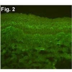
- Immunofluorescent analysis of bovine corneal epithelium (CE) limbus tissue extracts using Calcium Sensing Receptor Monoclonal Antibody (Product # MA1-934).

- Submitted by
- Invitrogen Antibodies (provider)
- Main image
- Experimental details
- Immunohistochemical analysis of mouse stomach stained with Calcium Sensing Receptor Monoclonal Antibody (Product # MA1-934). Fresh stomach tissue was fixed in 4% formalin for 1 hour and then incubated overnight at 4°C in 25% sucrose before embedding in tissue freezing medium. Antigen retrieval was carried out on 8 µm cryo-sections by incubating in sodium citrate buffer for 45 minutes at 4°C, immersing in sodium citrate buffer for 10 minutes at 100°C before washing 3 times for 5 minutes each in 1X PBS. Sections were then blocked in blocking buffer (0.3% Triton X-100 in 1X PBS containing 10% normal goat serum) for 30 minutes at RT before staining with Product # MA1-934 (diluted 1:100 in blocking buffer) overnight at 4°C followed by a fluorophore-conjugated goat anti-mouse IgG secondary antibody for 2 hours at RT. Sections were also stained with DAPI nuclear stain (blue). Product # MA1-934 positive cells (green) were found at the base of the antral glands in the mouse stomach. Data courtesy of the Innovators Program.

- Submitted by
- Invitrogen Antibodies (provider)
- Main image
- Experimental details
- Immunohistochemistry was performed on normal biopsies of deparaffinized Human brain tissue. To expose target proteins, heat induced antigen retrieval was performed using 10mM sodium citrate (pH6.0) buffer, microwaved for 8-15 minutes. Following antigen retrieval tissues were blocked in 3% BSA-PBS for 30 minutes at room temperature. Tissues were then probed at a dilution of 1:100 with a mouse monoclonal antibody recognizing Calcium Sensing Receptor (Product # MA1-934) or without primary antibody (negative control) overnight at 4°C in a humidified chamber. Tissues were washed extensively with PBST and endogenous peroxidase activity was quenched with a peroxidase suppressor. Detection was performed using a biotin-conjugated secondary antibody and SA-HRP, followed by colorimetric detection using DAB. Tissues were counterstained with hematoxylin and prepped for mounting.
Supportive validation
- Submitted by
- Invitrogen Antibodies (provider)
- Main image
- Experimental details
- NULL
- Submitted by
- Invitrogen Antibodies (provider)
- Main image
- Experimental details
- NULL
- Submitted by
- Invitrogen Antibodies (provider)
- Main image
- Experimental details
- NULL

- Submitted by
- Invitrogen Antibodies (provider)
- Main image

- Experimental details
- NULL
- Submitted by
- Invitrogen Antibodies (provider)
- Main image
- Experimental details
- NULL
- Submitted by
- Invitrogen Antibodies (provider)
- Main image
- Experimental details
- NULL
- Submitted by
- Invitrogen Antibodies (provider)
- Main image
- Experimental details
- NULL

- Submitted by
- Invitrogen Antibodies (provider)
- Main image
- Experimental details
- NULL
- Submitted by
- Invitrogen Antibodies (provider)
- Main image
- Experimental details
- NULL

- Submitted by
- Invitrogen Antibodies (provider)
- Main image

- Experimental details
- NULL
- Submitted by
- Invitrogen Antibodies (provider)
- Main image
- Experimental details
- NULL

- Submitted by
- Invitrogen Antibodies (provider)
- Main image
- Experimental details
- NULL

- Submitted by
- Invitrogen Antibodies (provider)
- Main image
- Experimental details
- NULL
- Submitted by
- Invitrogen Antibodies (provider)
- Main image
- Experimental details
- NULL
- Submitted by
- Invitrogen Antibodies (provider)
- Main image
- Experimental details
- NULL
- Submitted by
- Invitrogen Antibodies (provider)
- Main image
- Experimental details
- NULL
- Submitted by
- Invitrogen Antibodies (provider)
- Main image

- Experimental details
- NULL
- Submitted by
- Invitrogen Antibodies (provider)
- Main image

- Experimental details
- NULL

- Submitted by
- Invitrogen Antibodies (provider)
- Main image
- Experimental details
- NULL
- Submitted by
- Invitrogen Antibodies (provider)
- Main image
- Experimental details
- NULL
- Submitted by
- Invitrogen Antibodies (provider)
- Main image
- Experimental details
- NULL
- Submitted by
- Invitrogen Antibodies (provider)
- Main image
- Experimental details
- NULL

- Submitted by
- Invitrogen Antibodies (provider)
- Main image
- Experimental details
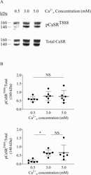
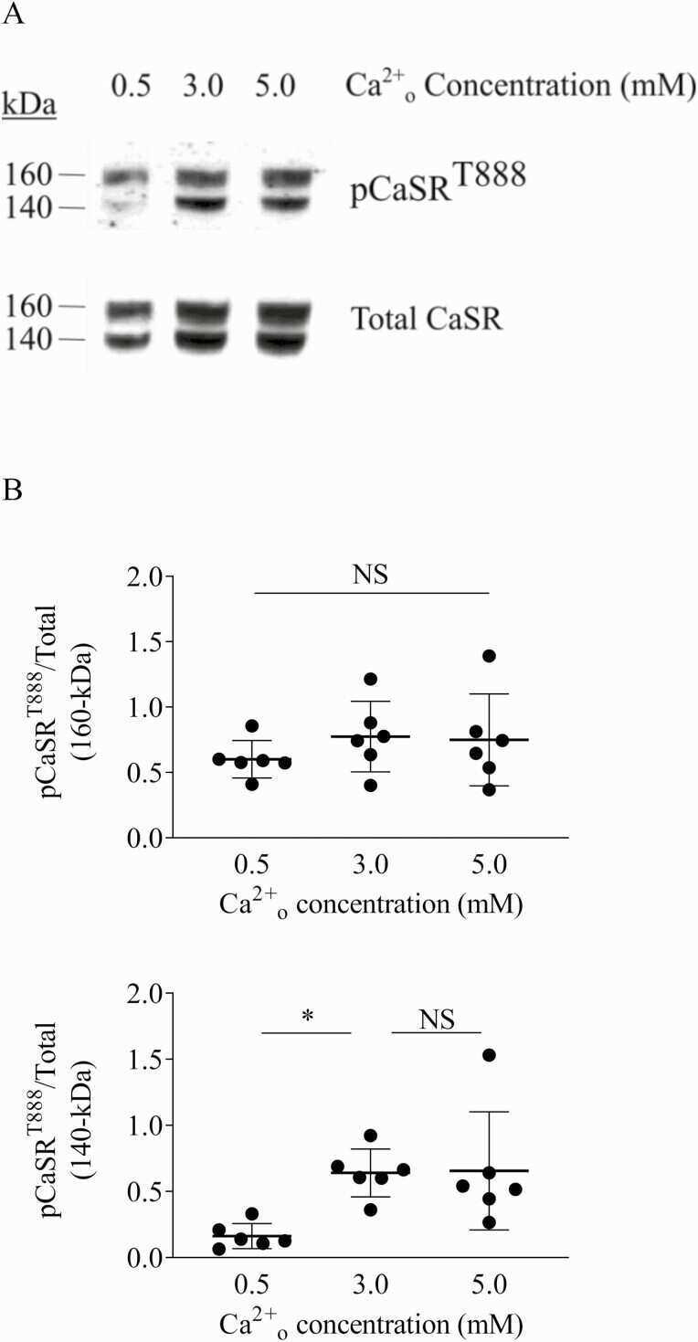
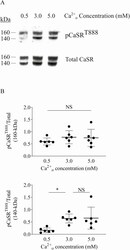
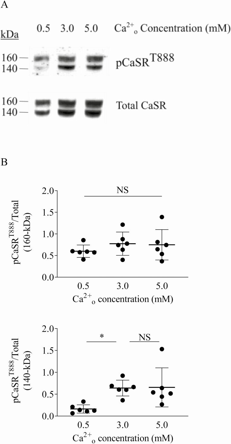
- Figure 6. Impact of Ca 2+ o concentration on phosphorylation of CaSR residue T888. (A) Detection and quantitation of CaSR T888 phosphorylation, and total CaSR in HEK-CaSR cells that were incubated in 0.2% BSA supplemented DMEM at 0.5, 3.0, or 5.0mM Ca 2+ o for 24 hours, followed by Western blotting. Over the extended period of the experiment, we did not observe high Ca 2+ o -induced dephosphorylation of CaSR-T888. Representative Western blot from 6 independent experiments. (B) Quantitation of Western blot intensity data. In response to either 3.0mM or 5.0mM Ca 2+ o a stable increase in pCaSR T888 was observed for the 140 kDa (high mannose) form but not the 160 kDa (complex glycosylated) form.
- Submitted by
- Invitrogen Antibodies (provider)
- Main image
- Experimental details
- NULL

- Submitted by
- Invitrogen Antibodies (provider)
- Main image
- Experimental details
- NULL
- Submitted by
- Invitrogen Antibodies (provider)
- Main image
- Experimental details
- Figure 1 CaSR protein expression in HUVECs by Immunofluorescence Confocal Microscopy and Western Blot. Immunofluorescent localization of CaSR in HUVECs with specific antibody and negative control after fixation and permeabilization protocol (A and B), or after fixation but not membrane permeabilization (C and D). Representative Western Blot of CaSR protein levels in HAoVSMC, HAEC and HUVEC total lysate, and in HUVEC membrane and cytoplasm extracts (E).

- Submitted by
- Invitrogen Antibodies (provider)
- Main image
- Experimental details
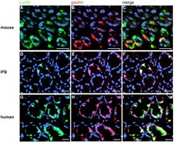
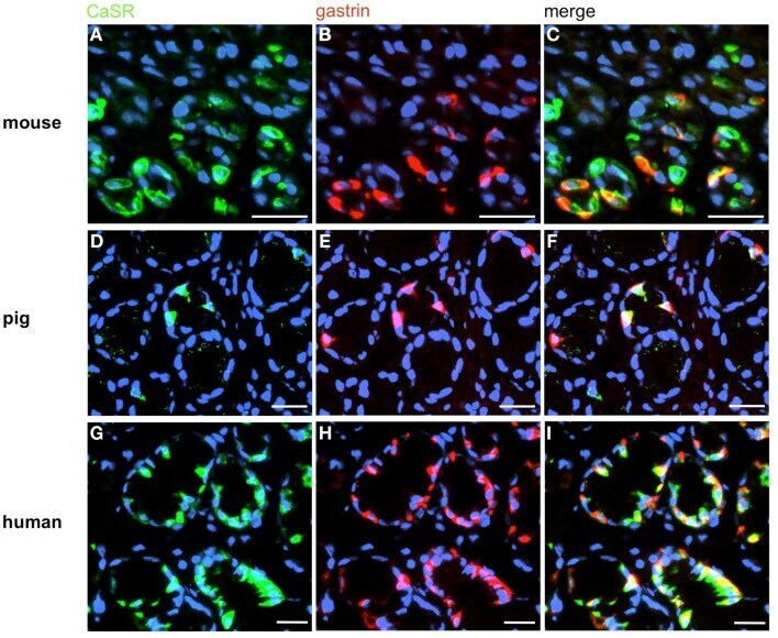
- Figure 5 Visualization of CaSR in murine, porcine, and human G-cells . Double-immunolabeling of horizontal tissue sections through the antral mucosa of mouse, swine and men for CaSR (green), and gastrin (red). (A,D,G) The CaSR antibody labels numerous cells in the antrum mucosa of mouse (A) , pig (D) , and men (G) . (B,E,H) Gastrin-positive cells are located in the same antral regions. (C,F,I) Overlay of accordant images; murine (C) , porcine (F) , and human (I) CaSR-positive cells are also immunoreactive for gastrin. Sections are counterstained with DAPI (blue). Scale bars: (A-I) = 20 mum.
- Submitted by
- Invitrogen Antibodies (provider)
- Main image
- Experimental details
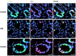
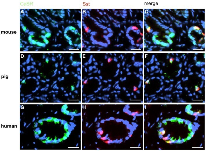
- Figure 7 A small subpopulation of CaSR-positive cells contains somatostatin (Sst) . Double-label immunohistochemistry employing antibodies for CaSR (green) and Sst (red) on horizontal tissue sections through the antrum of mouse, pig, and men. (A,D,G) The CaSR antibody labels numerous cells in the antrum mucosa of mouse (A) , pig (D) , and men (G) . (B,E,H) In the same annuli numerous Sst-positive cells can be visualized. (C,F,I) Overlay of accordant images; subpopulations of murine (C) , porcine (F) , and human (I) CaSR-positive cells show immunoreactivity for somatostatin. Sections are counterstained with DAPI (blue). Scale bars: (A-I) = 20 mum.
- Submitted by
- Invitrogen Antibodies (provider)
- Main image
- Experimental details
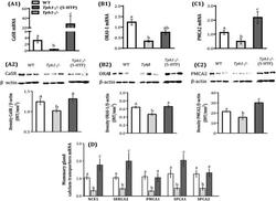
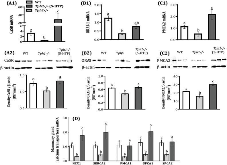
- Figure 3 Serotonin deficiency decreases calcium transporters expression in the mammary gland on d 10 of lactation. Wild-type (WT) mouse dams were given saline and Tryptophan hydroxylase knock-out dams given either saline ( Tph1 -/- ) or daily injections of exogenous 5-HTP (100 mg/kg; Tph1 -/- (5-HTP)), from pregnancy d20 to lactation d10. Mammary gland mRNA and protein expression of calcium sensing receptor (CaSR, A1-2), calcium release-activated calcium channel protein 1 (ORAI-1, B1-2) and plasma membrane Ca 2+ ATPase and 2 (PMCA2, C1-2) on d10 of lactation. (D) Mammary mRNA expression of sodium/calcium exchanger 1 (NCX1), sarcoplasmic endoplasmic Ca 2+ ATPase 2 (SERCA2), plasma membrane Ca 2+ ATPase 1 (PMCA1), secretory pathway Ca 2+ ATPase 1 and 2 (SPCA1 and 2) on d10 of lactation. All values are the mean +- SEM (n = 7). Groups with different letters are significantly different ( P
- Submitted by
- Invitrogen Antibodies (provider)
- Main image
- Experimental details
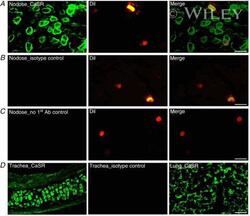
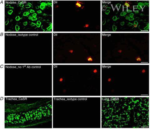
- 1 Expression of calcium-sensing receptor (CaSR) in rat vagal bronchopulmonary sensory neurons, trachea and lung parenchyma A , CaSR immunostaining (green) in a cryosection of nodose ganglion. B , negative control with primary antiserum for CaSR substituted by its isotype, normal mouse IgG2a, in a nodose section. C , negative control with primary antiserum (Ab) for CaSR omitted in a nodose section. 1,1'-Dioctadecyl-3,3,3',3'-tetramethylindocarbocyanine perchlorate (DiI) labelling (red) shown in the middle and right panels in A - C identifies the sensory neurons innervating the airway and lung structures. D , left panel, CaSR staining in trachea; middle panel, isotype control for CaSR in trachea; and right panel, CaSR staining in lung parenchyma. All tissues were sectioned at 12 mum; scale bars represent 50 mum.
- Submitted by
- Invitrogen Antibodies (provider)
- Main image
- Experimental details
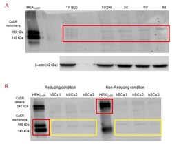
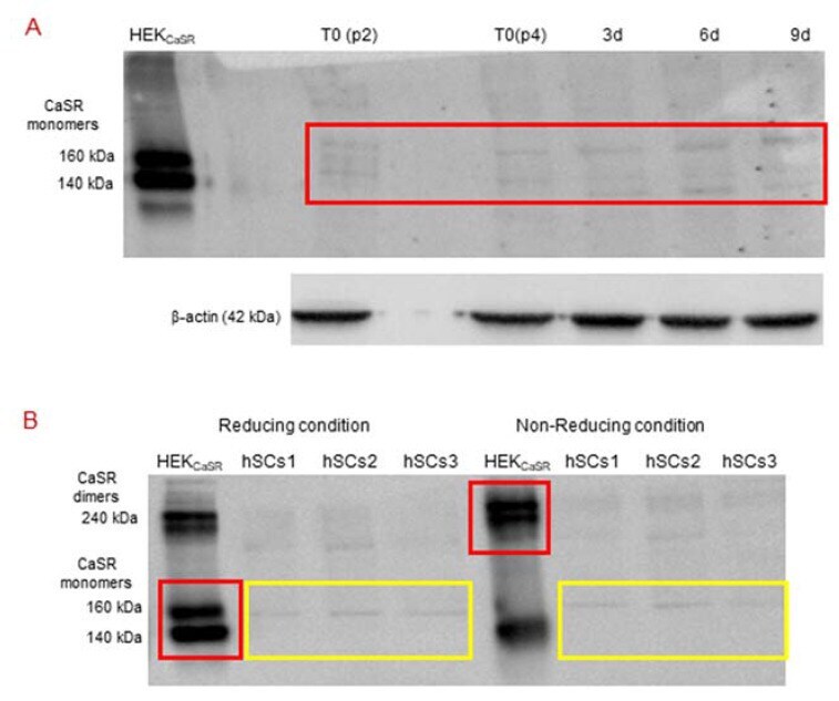
- Figure 5 ( A ) Western Blot (standard reducing condition) for CaSR protein in hSC lines and during myogenic differentiation. The results show the presence of faint bands of about 160 kDa and 140 kDa in hSC lines and at different time points (T 0 , 3, 6, 9 days of myogenic differentiation) of primary hSC lines. HEK CaSR were used as positive control. Experiments were carried out in triplicate and are representative of the three established hSC lines. ( B ) Western blot analysis of CaSR in reducing and non-reducing conditions. The analysis for CaSR protein in three hSC lines showed that the observed bands are not CaSR as they did not shift to dimer form at 240 kDa in non-reducing conditions. HEK CaSR were used as positive control. The red and yellow boxes surround the analyzed bands.
- Submitted by
- Invitrogen Antibodies (provider)
- Main image

- Experimental details
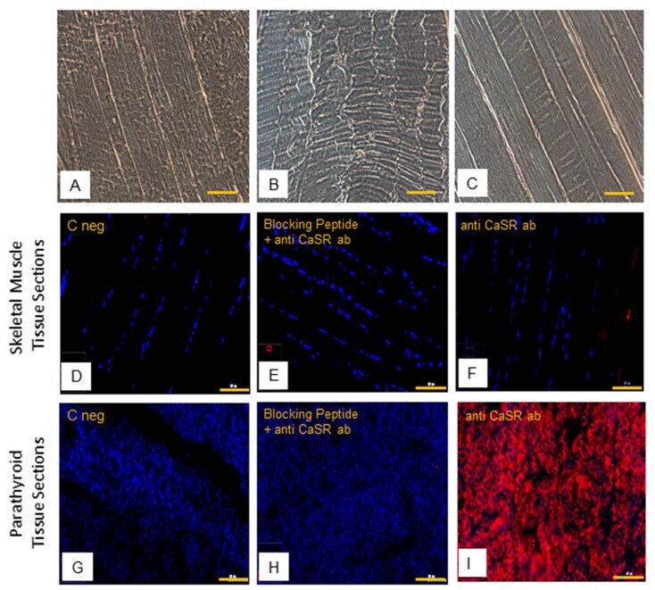
- Figure 6 Human skeletal muscle tissue (hSMt) sections; observations in phase contrast microscopy, original. Magnification: 10x, scale bar 200 um ( A - C ). Immunostaining of CaSR protein. Analysis of CaSR protein in human skeletal muscle tissue sections: ( D ) negative control only with secondary antibody, ( E ) control with anti-CaSR antibody absorbed with blocking peptide, and ( F ) with primary anti-CaSR antibody. Analysis of CaSR protein in human parathyroid tissue sections used as positive control: ( G ) negative control with only secondary antibody, ( H ) control with anti-CaSR antibody absorbed with blocking peptide, and ( I ) with primary anti-CaSR antibody. Fluorescent microscopy in conventional colors: red for CaSR and blue for nuclei, original magnification: 10x, scale bar: 200 um. Experiment was carried out in triplicate (in hSMt sections of three different humans).
- Submitted by
- Invitrogen Antibodies (provider)
- Main image
- Experimental details
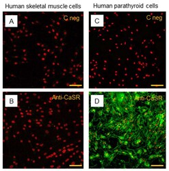
- Figure 7 Immunostaining of CaSR in hSC lines. The analysis of CaSR protein in hSC lines: ( A ) negative control with only secondary antibody and ( B ) with primary anti-CaSR antibody. Results are representative of experiments carried out in three established hSC lines. The analysis of CaSR protein was also performed in human parathyroid cells, used as positive control, ( C ) negative control with only secondary antibody, and ( D ) with primary anti-CaSR antibody. LSCM conventional colors: green for CaSR protein and red for nuclei, original magnification: 20x, scale bar: 50 um.
- Submitted by
- Invitrogen Antibodies (provider)
- Main image
- Experimental details
- Figure 6. Impact of Ca 2+ o concentration on phosphorylation of CaSR residue T888. (A) Detection and quantitation of CaSR T888 phosphorylation, and total CaSR in HEK-CaSR cells that were incubated in 0.2% BSA supplemented DMEM at 0.5, 3.0, or 5.0mM Ca 2+ o for 24 hours, followed by Western blotting. Over the extended period of the experiment, we did not observe high Ca 2+ o -induced dephosphorylation of CaSR-T888. Representative Western blot from 6 independent experiments. (B) Quantitation of Western blot intensity data. In response to either 3.0mM or 5.0mM Ca 2+ o a stable increase in pCaSR T888 was observed for the 140 kDa (high mannose) form but not the 160 kDa (complex glycosylated) form.
- Submitted by
- Invitrogen Antibodies (provider)
- Main image
- Experimental details
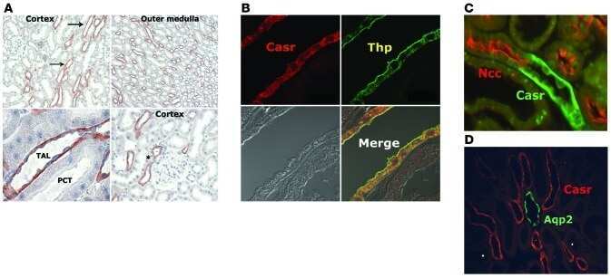
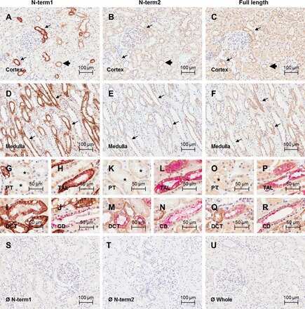
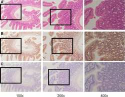
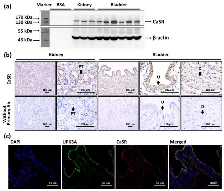
- Figure 1 The expression and location of calcium-sensing receptor (CaSR) in bladder. ( a ) Western blot analysis of CaSR (MW: 130 kDa) in the whole kidney and whole urinary bladder. ( b ) Immunohistochemistry staining of CaSR (brown signals) in the urothelium (U) and detrusor (D) of bladder, and in the proximal tubule (PT) of kidney, and the staining without applying primary antibody (Ab). ( c ) Immunofluorescence staining of CaSR (red), uroplakin III A (UPK3A) (green), and DAPI (blue) in urothelium of urinary bladder.
- Submitted by
- Invitrogen Antibodies (provider)
- Main image
- Experimental details
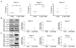
- Figure 2 Establishment of the PTH-secretion potential of T-MSC-PTHCs from three donors: ( A ) conditioned medium was taken each step of T-MSC and T-MSC-PTHCs, and the concentration of PTH was measured using a commercial PTH ELISA kit at high cell density. Data are presented as the mean +- SD of at least three experiments (* p < 0.05, ** p < 0.01). PTH concentrations were significantly increased in T-MSC-PTHCs, compared with T-MSCs derived from donors #1 and #2, respectively. The expression of CaSR protein during differentiation of T-MSCs into T-MSC-PTHCs was measured by Western blotting and quantified using ImageJ software at low ( B ) and high ( C ) cell densities. Protein levels are normalized to GAPDH. Data are presented as the mean +- SD of at least three experiments (*** p < 0.001). CaSR expression levels were significantly increased in T-MSC-PTHCs, compared with T-MSCs derived from donors #1 and #3, respectively. Abbreviations: PTH, parathyroid hormone; T-MSC, tonsil-derived mesenchymal stem cells; T-MSC-PTHCs, T-MSC-derived PTH-releasing cells; CaSR, calcium-sensing receptor: GAPDH, glyceraldehyde 3-phosphate dehydrogenase.
- Submitted by
- Invitrogen Antibodies (provider)
- Main image
- Experimental details
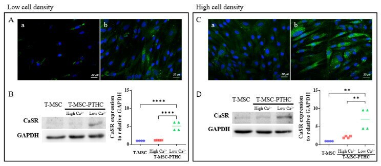
- Figure 3 Comparison of differentiation potential of T-MSC-PTHCs according to cell density during differentiation (low cell density: A, B; high cell density: ( C , D )): ( A , C ) the expression of PTH (green) in T-MSC (panel a) and T-MSC-PTHCs (panel b) derived from donor #1 was evaluated by immunostaining. The cells were counterstained with DAPI (blue). The expression of PTH of T-MSC-PTHCs was higher in high-density cells ( C ) than low-density cells ( A ); ( B , D ) expression of CaSR protein according to extracellular calcium level in T-MSC-PTHCs from three donor #1. T-MSC-PTHCs were further exposed to culture media containing low (0.09 mM) or high (3.0 mM) calcium concentrations for up to 48 h. The expression of CaSR was measured by Western blotting and quantified using ImageJ software and are normalized to GAPDH. Data are presented as the mean +- SD of at least three experiments (** p < 0.01, **** p < 0.0001). In both densities of cells, the expression of CaSR protein was significantly increased under exposure to low calcium and was not significantly increased under exposure to high calcium. Abbreviations: PTH, parathyroid hormone; T-MSC, tonsil-derived mesenchymal stem cells; T-MSC-PTHCs, T-MSC-derived PTH-releasing cells; CaSR, calcium-sensing receptor; GAPDH, glyceraldehyde 3-phosphate dehydrogenase.
- Submitted by
- Invitrogen Antibodies (provider)
- Main image
- Experimental details
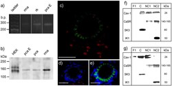
- Figure 6. Immunolocalization and identification of calcium-sensing receptor (CaSR) protein and mRNA. a, Amplicons of the anticipated size (283 bp) were produced by RT-PCR amplification of RNA obtained from rat mesenteric artery (rma) and kidney (rk) and from porcine coronary artery endothelial cells (pca E). b, Western blot analysis of homogenates obtained from HEK293 cells transfected with CaSR (HEK), rat mesenteric and porcine coronary arteries (pca), and porcine coronary artery endothelial cell samples (each 35 &mgrg protein) using mouse monoclonal anti-CaSR antibodies. c and d, Same transverse section of rat mesenteric artery without (c) or with (d) DAPI staining of nuclei (blue). Immunoreactivity to the anti-CaSR antibody (red) was observed in the single layer of endothelial cells which is separated from the multiple layers of myocytes by the internal elastic lamina (green autofluorescence). The external elastic lamina defines the inner limit of the adventitial layer in which strong immunoreactivity to the antibody was also observed. e, Section of artery incubated with DAPI and secondary, but not primary, antibody. In (c through e), the horizontal scale bar represents 100 &mgrm. After density gradient separation of membrane fractions from (f) rat mesenteric artery and (g) porcine coronary artery endothelial cells, SK3 immunoreactivity was associated with the caveolin-1-rich fraction (C) whereas IK1 and CaSR were predominantly associated with the less buoyant (NC2) of the
- Submitted by
- Invitrogen Antibodies (provider)
- Main image
- Experimental details
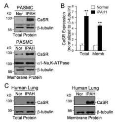
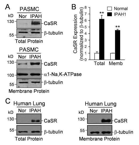
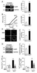
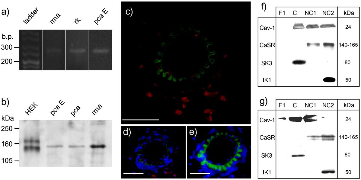
- Figure 2. Upregulation of CaSR in PASMC from IPAH patients. A , Western blot analysis on CaSR in total proteins ( top ) and membrane proteins ( bottom ) isolated from normal PASMC (Nor) and IPAH-PASMC (IPAH). B , Summarized data (mean+-SE), bottom showing CaSR protein levels that were normalized to the &bgr-tubulin level. &ast&ast P

- Submitted by
- Invitrogen Antibodies (provider)
- Main image

- Experimental details
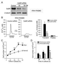
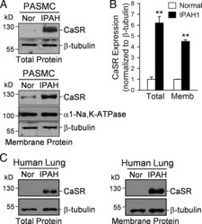
- Figure 4. Downregulation of CaSR with siRNA attenuates extracellular Ca 2&plus -induced &lsqbCa 2&plus &rsqb cyt increases in IPAH-PASMC and inhibits IPAH-PASMC proliferation. A , Western blot analysis on CaSR in IPAH-PASMC treated with a control (or scrambled) siRNA (control) and siRNA for CaSR (at 25 and 50 nmol&solL, n=3 for each group). B , Representative records of &lsqbCa 2&plus &rsqb cyt changes ( left ) and summarized data (mean+-SE) ( right ) showing the extracellular Ca 2&plus -induced &lsqbCa 2&plus &rsqb cyt increases in IPAH-PASMC transfected with control siRNA (n=57) and CaSR-siRNA (n=57). &ast&ast P
- Submitted by
- Invitrogen Antibodies (provider)
- Main image

- Experimental details
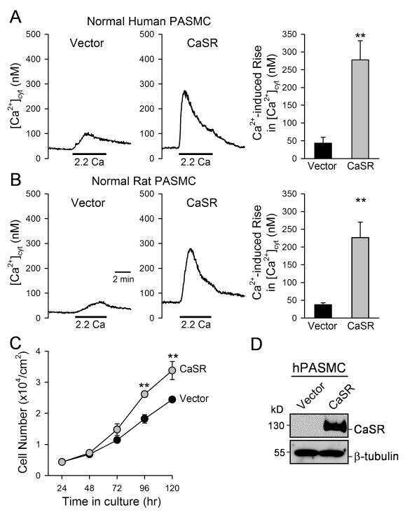
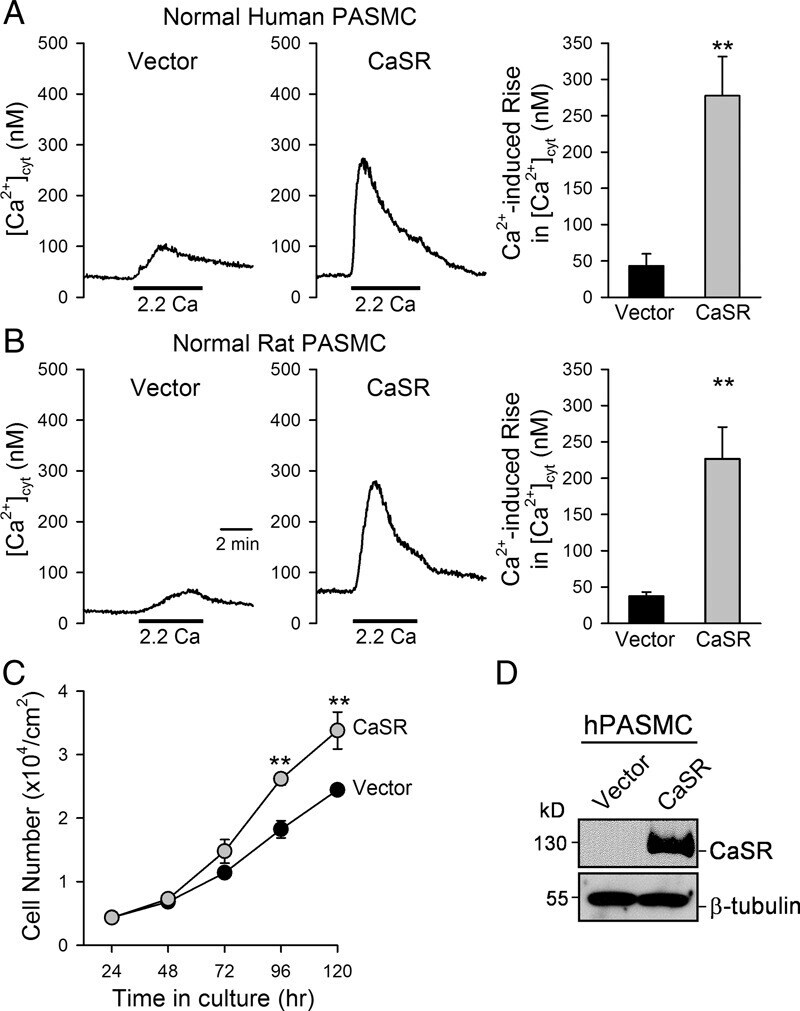
- Figure 5. Overexpression of CaSR in normal PASMC enhances extracellular Ca 2&plus -induced &lsqbCa 2&plus &rsqb cyt increase and promotes cell proliferation. A and B , Representative records of &lsqbCa 2&plus &rsqb cyt changes ( left ) and summarized data (mean+-SE) ( right ) extracellular Ca 2&plus -induced &lsqbCa 2&plus &rsqb cyt increases in human ( A ) and rat ( B ) PASMC transfected with an empty vector (vector, n=12) and the human CaSR cDNA (CaSR, n=8). C , Summarized data (mean+-SE) showing the total numbers of normal human PASMC transfected with the vector (solid circles) and CaSR gene (gray circles) after incubation in growth media for 24, 48, 72, 96, and 120 hours. The growth curves for vector control PASMC and IPAH PASMC are significantly different ( P
- Submitted by
- Invitrogen Antibodies (provider)
- Main image

- Experimental details
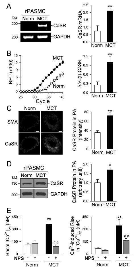
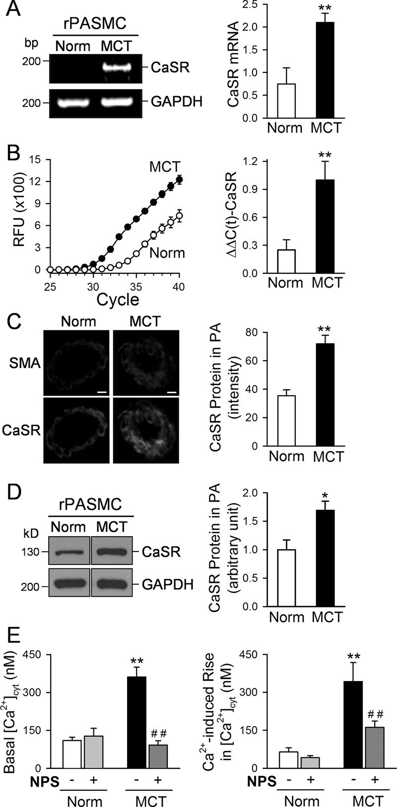
- Figure 6. Upregulation of CaSR in PASMC from rats with MCT-induced pulmonary hypertension. A and B , Regular ( A ) and real-timer ( B ) RT-PCR analyses on CaSR in PASMC isolated from normal rats (Norm, n=6) and rats with MCT-induced pulmonary hypertension (MCT, n=5). &ast&ast P
- Submitted by
- Invitrogen Antibodies (provider)
- Main image
- Experimental details
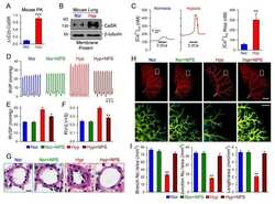
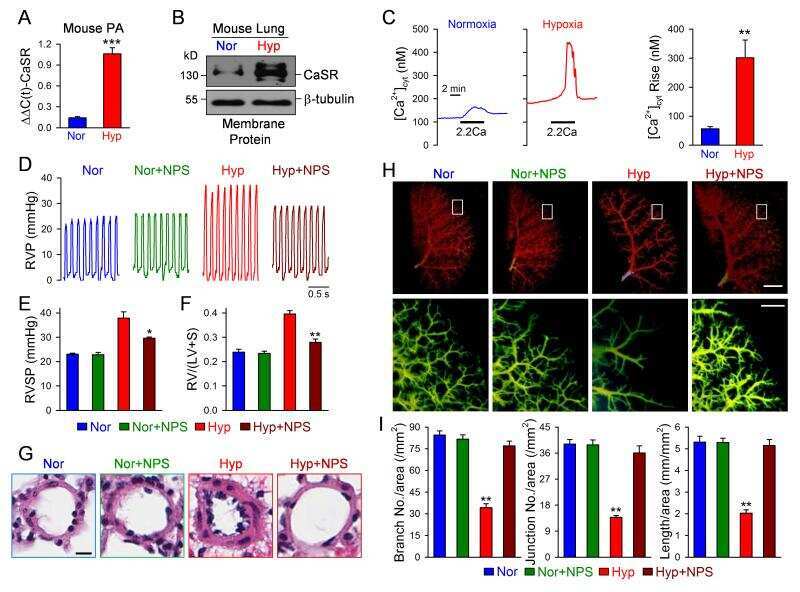
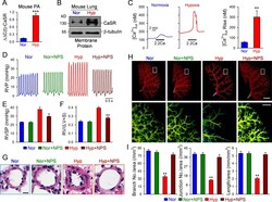
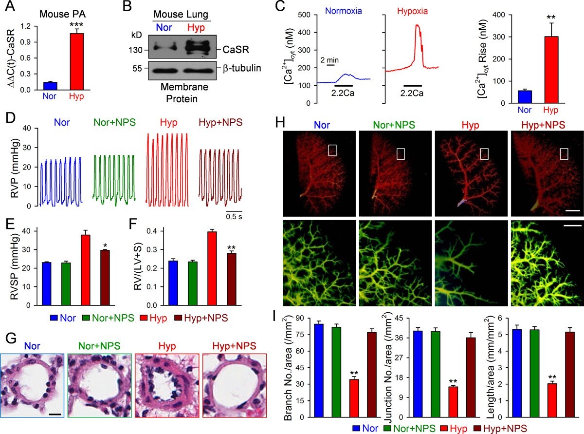
- Figure 8. Upregulation of CaSR in PASMC from HPH mice and blockade of CaSR by NPS 2143 inhibits the development of HPH in mice. A and B , Real-time RT-PCR ( A ) and Western blot ( B ) analyses on CaSR in PA and lung tissues isolated from normoxic (Nor) and hypoxic (Hyp) mice. C , Representative record ( left ) and summarized data (mean+-SE, right ) showing the extracellular Ca 2&plus -induced increase in &lsqbCa 2&plus &rsqb cyt in PASMC isolated from normoxic and hypoxic mice. &ast&ast P
- Submitted by
- Invitrogen Antibodies (provider)
- Main image
- Experimental details
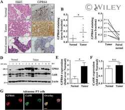
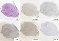
- Figure 1. CYP11B1, CYP11B2, PTH1R, CaSR, and VD3R immunohistochemical expressions of irregular cord-like distribution in case 1.

- Submitted by
- Invitrogen Antibodies (provider)
- Main image

- Experimental details
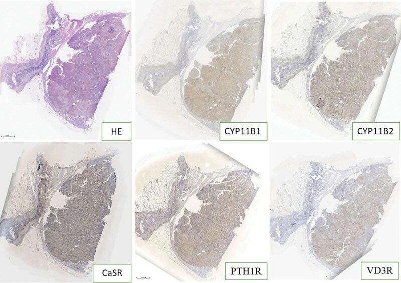
- Figure 2. CYP11B1, CYP11B2, PTH1R, CaSR, and VD3R immunohistochemical expressions of multiple germinal centers in case 2.
- Submitted by
- Invitrogen Antibodies (provider)
- Main image
- Experimental details
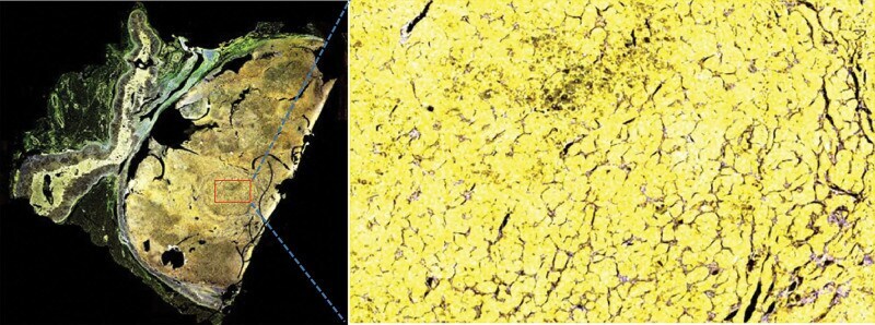
- Figure 5. CYP11B1, CYP11B2, PTH1R, CaSR, and VD3R multiple fluorescence immunohistochemistry with overlapping distribution areas in case 2.
- Submitted by
- Invitrogen Antibodies (provider)
- Main image

- Experimental details
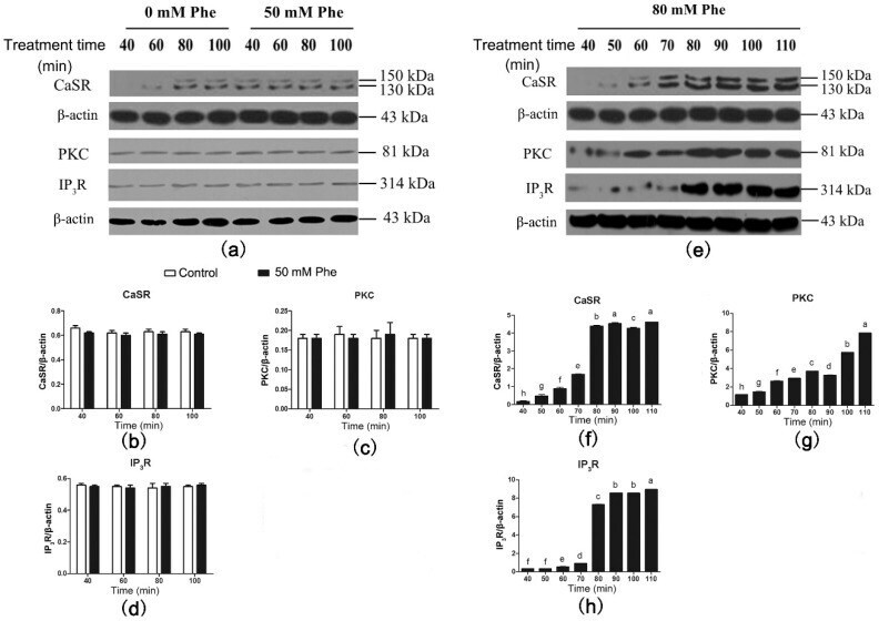
- Figure 5 Effect of Phe (0 mM, 50 mM, and 80 mM) on the levels of CaSR-mediated downstream pathway in the pig duodenum. The perfusion of duodenal tissues were done with 0 (control group), 50 mM, and 80 mM Phe for 160 min, respectively. During the perfusion, the perfused tissues were recovered every 10 min, starting from 40 min of perfusion to 110 min of perfusion, to extract total protein. ( a , e ) The measurement of protein levels of ( b , f ) CaSR, ( c , g ) PKC, and ( d , h ) IP 3 R in the duodenum tissues was performed by western blot analysis, using beta-actin as a loading control. Data are expressed as mean +- SEM ( n = 3). Dissimilar letters represent a significant difference at p < 0.05.
- Submitted by
- Invitrogen Antibodies (provider)
- Main image
- Experimental details
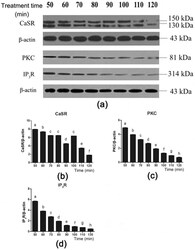
- Figure 6 Effect of the CaSR antagonist NPS 2143 on the CaSR-mediated downstream pathway in the presence of 80 mM Phe in pig duodenum. The tissues were perfused with 80 mM Phe and 25 uM NPS 2143 (specific CaSR antagonist) for 160 min. During the perfusion, the perfused tissues were recovered every 10 min, starting from 50 min to 120 min, to extract total protein. ( a ) The measurement of protein levels of ( b ) CaSR, ( c ) PKC, and ( d ) IP 3 R in the duodenum tissues was performed by western blot analysis, using beta-actin as a loading control. Data are expressed as mean +- SEM ( n = 3). Dissimilar letters represent a significant difference at p < 0.05.

- Submitted by
- Invitrogen Antibodies (provider)
- Main image

- Experimental details
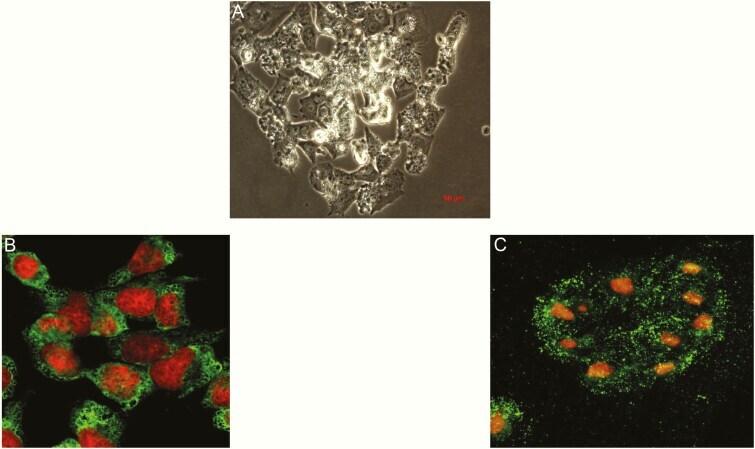
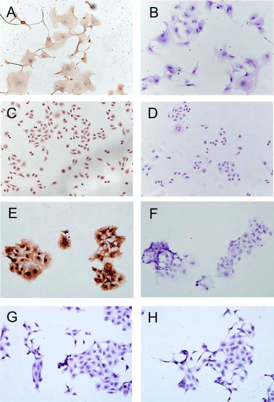
- Figure 1. The intracellular PTH and CaSR proteins were evaluated with immunofluorescence staining in adenoma parathyroid cells after 5 days in culture with growth medium and then transferred into glass slides and incubated for 24 h in DMEM medium containing 1.2 mM calcium before the phase contrast analysis and the immunofluorescence staining. (A) Phase contrast image of an adenoma parathyroid primary culture (40x original magnification). (B) Parathyroid cells stained for CaSR (in green) and counterstained for nuclei (in red). Observations with laser scanning confocal microscopy, objective 40x original magnification. (C) Parathyroid cells stained for PTH (in green) and counterstained for nuclei (in red). Observations with laser scanning confocal microscopy, 40x original magnification.