Explore
 Validate
 Learn
Learn Western blot
 ELISA
ELISAAntibody data
- Antibody Data
- Antigen structure
- References [1]
- Comments [0]
- Validations
- Western blot [1]
- Immunocytochemistry [2]
- Immunohistochemistry [1]
Submit
Validation data
Reference
Comment
Report error
- Product number
- SM5143P - Provider product page

- Provider
- Acris Antibodies GmbH
- Proper citation
- Acris Antibodies GmbH Cat#SM5143P, RRID:AB_973558
- Product name
- anti CaSR
- Antibody type
- Monoclonal
- Antigen
- Synthetic peptide corresponding to amino acids 214-235 of Human Calcium Sensing Receptor (CaSR).
- Reactivity
- Human, Mouse, Rat, Bovine
- Host
- Mouse
- Isotype
- IgG
- Antibody clone number
- 5C10, ADD
- Vial size
- 0.1 mg
- Concentration
- 1.0 mg/ml
Submitted references Novel inactivating mutations of the calcium-sensing receptor: the calcimimetic NPS R-568 improves signal transduction of mutant receptors.
Rus R, Haag C, Bumke-Vogt C, Bähr V, Mayr B, Möhlig M, Schulze E, Frank-Raue K, Raue F, Schöfl C
The Journal of clinical endocrinology and metabolism 2008 Dec;93(12):4797-803
The Journal of clinical endocrinology and metabolism 2008 Dec;93(12):4797-803
No comments: Submit comment
Supportive validation
- Submitted by
- Acris Antibodies GmbH (provider)
- Main image

- Experimental details
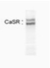
- Western blot of calcium sensing receptor in HEK7-2 cell extract using SM5143P.
Supportive validation
- Submitted by
- Acris Antibodies GmbH (provider)
- Main image
- Experimental details
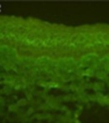
- Immunofluorescence of Bovine CE (Corneal Epithelium) Limbus tissue extracts using SM5143P.

- Submitted by
- Acris Antibodies GmbH (provider)
- Main image
- Experimental details
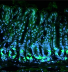
- Immunohistochemical analysis of mouse stomach stained with Calcium Sensing Receptor Monoclonal Antibody (Cat.-No SM5143P). Fresh stomach tissue was fixed in 4% formalin for 1 hour and then incubated overnight at 4ºC in 25% sucrose before embedding in tissue freezing medium. Antigen retrieval was carried out on 8µm cryo-sections by incubating in sodium citrate buffer for 45 minutes at 4ºC, immersing in sodium citrate buffer for 10 minutes at 100ºC before washing 3 times for 5 minutes each in 1X PBS. Sections were then blocked in blocking buffer (0.3% Triton X-100 in 1X PBS containing 10% normal goat serum) for 30 minutes at RT before staining with SM5143P (diluted 1:100 in blocking buffer) overnight at 4ºC followed by a fluorophore-conjugated goat anti-mouse IgG secondary antibody for 2 hours at RT. Sections were also stained with DAPI nuclear stain (blue). SM5143P positive cells (green) were found at the base of the antral glands in the mouse stomach. Data courtesy of the Innovators Program.

Supportive validation
- Submitted by
- Acris Antibodies GmbH (provider)
- Main image
- Experimental details
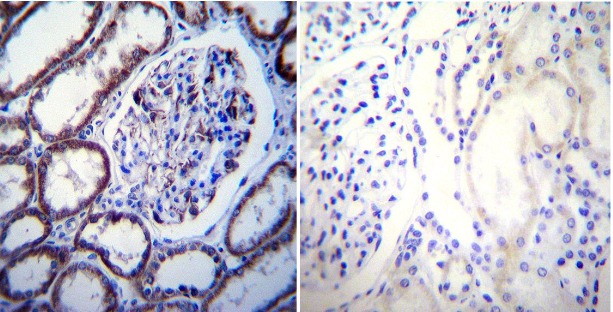
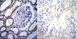
- Immunohistochemistry performed on normal biopsies of deparaffinized Human kidney tissue using CaSR Antibody Cat.-No SM5143P (Clone 5C10, ADD)