Explore
 Validate
 Learn
Learn14-9688-80
antibody from Invitrogen Antibodies
Targeting: LMNA
CMD1A, HGPS, LGMD1B, LMN1, LMNL1, MADA, PRO1
 Western blot
 Immunocytochemistry
Antibody data
- Antibody Data
- Antigen structure
- References [5]
- Comments [0]
- Validations
- Immunocytochemistry [2]
- Flow cytometry [2]
- Other assay [2]
Submit
Validation data
Reference
Comment
Report error
- Product number
- 14-9688-80 - Provider product page

- Provider
- Invitrogen Antibodies
- Product name
- Lamin A/C Monoclonal Antibody (4A7), eBioscience™
- Antibody type
- Monoclonal
- Antigen
- Other
- Description
- The monoclonal antibody 4A7 recognizes human, mouse, rabbit and hamster lamin A/C. Lamins are nuclear intermediate filament proteins that provide framework for the nuclear envelope, maintain cell morphology, and protect the nucleus from mechanical, thermal, and oxidative stresses. Lamins also play a role in nuclear assembly, chromatin organization, DNA replication, RNA transcription, cell signaling, and apoptosis. Lamin C is a splice variant of Lamin A and both are A-type lamins. Lamin A and C are crucial for skeletal and cardiac development and function. Defects in A-type lamins result in cardiomyopathy, muscular dystrophy, peripheral neuropathy, lipodystrophy, restrictive dermopathy, and progeroid disorders. This 4A7 antibody has been tested by immunocytochemistry of methanol-fixed and permeabilized cells and can be used at less than or equal to 10 µg/mL. It is recommended that the antibody be carefully titrated for optimal performance in the assay of interest. Purity: Greater than 90%, as determined by SDS-PAGE. Aggregation: Less than 10%, as determined by HPLC. Filtration: 0.2 µm post-manufacturing filtered.
- Reactivity
- Human, Mouse, Hamster, Rabbit
- Host
- Mouse
- Isotype
- IgG
- Antibody clone number
- 4A7
- Vial size
- 25 µg
- Concentration
- 0.5 mg/mL
- Storage
- 4°C
Submitted references FMRP Control of Ribosome Translocation Promotes Chromatin Modifications and Alternative Splicing of Neuronal Genes Linked to Autism.
Nuclear lamins in the brain - new insights into function and regulation.
Lamins in development, tissue maintenance and stress.
Lamin A and ZMPSTE24 (FACE-1) defects cause nuclear disorganization and identify restrictive dermopathy as a lethal neonatal laminopathy.
A lamin A/C beta-strand containing the site of lipodystrophy mutations is a major surface epitope for a new panel of monoclonal antibodies.
Shah S, Molinaro G, Liu B, Wang R, Huber KM, Richter JD
Cell reports 2020 Mar 31;30(13):4459-4472.e6
Cell reports 2020 Mar 31;30(13):4459-4472.e6
Nuclear lamins in the brain - new insights into function and regulation.
Jung HJ, Lee JM, Yang SH, Young SG, Fong LG
Molecular neurobiology 2013 Feb;47(1):290-301
Molecular neurobiology 2013 Feb;47(1):290-301
Lamins in development, tissue maintenance and stress.
Zuela N, Bar DZ, Gruenbaum Y
EMBO reports 2012 Dec;13(12):1070-8
EMBO reports 2012 Dec;13(12):1070-8
Lamin A and ZMPSTE24 (FACE-1) defects cause nuclear disorganization and identify restrictive dermopathy as a lethal neonatal laminopathy.
Navarro CL, De Sandre-Giovannoli A, Bernard R, Boccaccio I, Boyer A, Geneviève D, Hadj-Rabia S, Gaudy-Marqueste C, Smitt HS, Vabres P, Faivre L, Verloes A, Van Essen T, Flori E, Hennekam R, Beemer FA, Laurent N, Le Merrer M, Cau P, Lévy N
Human molecular genetics 2004 Oct 15;13(20):2493-503
Human molecular genetics 2004 Oct 15;13(20):2493-503
A lamin A/C beta-strand containing the site of lipodystrophy mutations is a major surface epitope for a new panel of monoclonal antibodies.
Manilal S, Randles KN, Aunac C, Nguyen Mt, Morris GE
Biochimica et biophysica acta 2004 Mar 17;1671(1-3):87-92
Biochimica et biophysica acta 2004 Mar 17;1671(1-3):87-92
No comments: Submit comment
Supportive validation
- Submitted by
- Invitrogen Antibodies (provider)
- Main image
- Experimental details
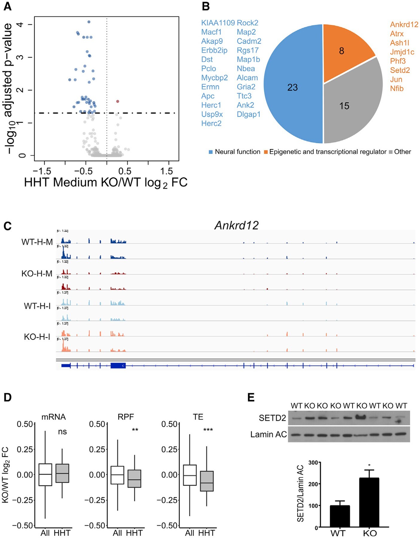
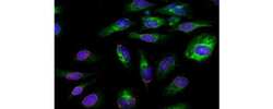
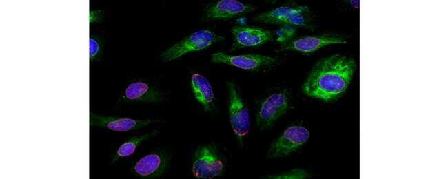
- Immunocytochemistry of fixed and permeabilized HeLa cells stained with 10 µg/mL of Anti-Lamin A/C Purified and 10 µg/mL of F(ab')2 Anti-Mouse IgG eFluor® 570 (red), followed by 1 µg/mL of Anti-Pan-Cytokeratin (AE1/AE3) Alexa Fluor® 488 (green).Nuclei are stained with DAPI (blue).
- Submitted by
- Invitrogen Antibodies (provider)
- Main image
- Experimental details
- Immunocytochemistry of fixed and permeabilized HeLa cells stained with 10 µg/mL of Anti-Lamin A/C Purified and 10 µg/mL of F(ab')2 Anti-Mouse IgG eFluor® 570 (red), followed by 1 µg/mL of Anti-Pan-Cytokeratin (AE1/AE3) Alexa Fluor® 488 (green).Nuclei are stained with DAPI (blue).

Supportive validation
- Submitted by
- Invitrogen Antibodies (provider)
- Main image

- Experimental details
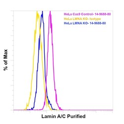
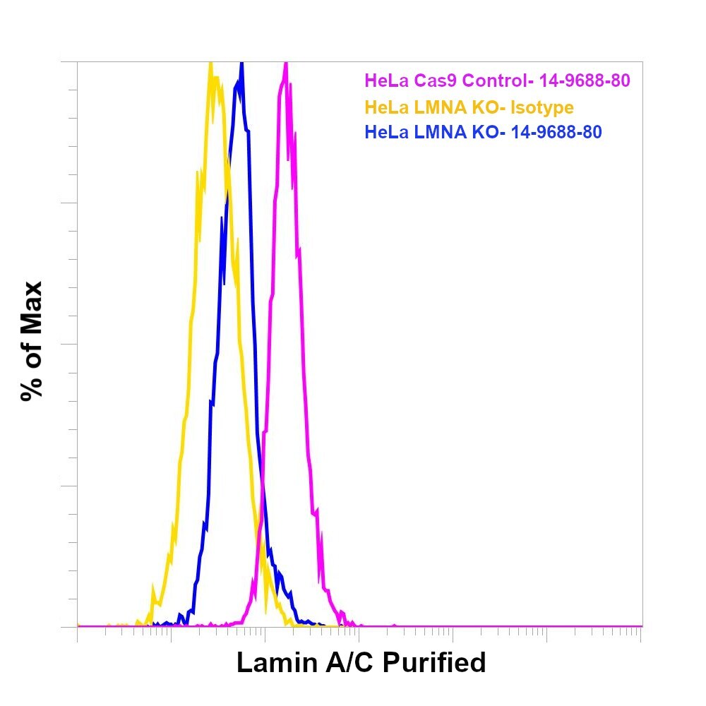
- Knockout of LMNA was achieved by CRISPR-Cas9 genome editing using LentiArray™ Lentiviral sgRNA (Product # A32042, Assay ID CRISPR817711_LV) and LentiArray Cas9 Lentivirus (Product # A32064). Flow cytometry analysis of Lamin A/C was performed by staining HeLa LMNA Knock out cells with10 µg/mL Mouse IgG1 kappa Isotype Control (P3.6.2.8.1), eBioscience™ (Product # 14-4714-82, yellow histogram) or10 µg/mLLamin A/C Monoclonal Antibody (4A7), eBioscience™ (Product # 14-9688-80, blue histogram) followed by Goat anti-Mouse IgG (H+L), Superclonal™ Recombinant Secondary Antibody, Alexa Fluor™ Plus 488 (Product # A55058, 1:1000).HeLa Cas9 control cells were also stained with10 µg/mLLamin A/C Monoclonal Antibody (4A7), eBioscience™ (Product # 14-9688-80, pink histogram) followed by the secondary antibody. Lossof signal was observed in the LMNA KOcells stained with LMNA antibody clone 4A7 but not in the control Cas9cells. Viable cells were used for analysis, as determined by Fixable Viability Dye eFluor™780 (Product # 65-0865-18).

- Submitted by
- Invitrogen Antibodies (provider)
- Main image
- Experimental details
- Knockout of LMNA was achieved by CRISPR-Cas9 genome editing using LentiArray™ Lentiviral sgRNA (Product # A32042, Assay ID CRISPR817711_LV) and LentiArray Cas9 Lentivirus (Product # A32064). Flow cytometry analysis of Lamin A/C was performed by staining HeLa LMNA Knock out cells with10 µg/mL Mouse IgG1 kappa Isotype Control (P3.6.2.8.1), eBioscience™ (Product # 14-4714-82, yellow histogram) or10 µg/mLLamin A/C Monoclonal Antibody (4A7), eBioscience™ (Product # 14-9688-80, blue histogram) followed by Goat anti-Mouse IgG (H+L), Superclonal™ Recombinant Secondary Antibody, Alexa Fluor™ Plus 488 (Product # A55058, 1:1000).HeLa Cas9 control cells were also stained with10 µg/mLLamin A/C Monoclonal Antibody (4A7), eBioscience™ (Product # 14-9688-80, pink histogram) followed by the secondary antibody. Lossof signal was observed in the LMNA KOcells stained with LMNA antibody clone 4A7 but not in the control Cas9cells. Viable cells were used for analysis, as determined by Fixable Viability Dye eFluor™780 (Product # 65-0865-18).
Supportive validation
- Submitted by
- Invitrogen Antibodies (provider)
- Main image
- Experimental details
- NULL

- Submitted by
- Invitrogen Antibodies (provider)
- Main image
- Experimental details
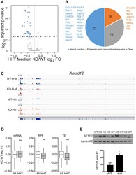
- Figure 5. FMRP Stalls Ribosomes on Specific mRNAs (A) RNA sedimenting to medium polysomes containing four to six ribosomes after HHT treatment of hippocampal slices; 46 RNAs are downregulated and 1 is upregulated in Fmr1 KO relative to WT. (B) Downregulated RNAs in HHT-treated Fmr1 KO slices primarily encode epigenetic and transcriptional regulators and proteins involved in neural function. (C) Example of Ankrd12 RNA, which has reduced reads in Fmr1 KO slices relative to WT after HHT (H) treatment. Input (I) reads are similar in both genotypes. M refers to medium fraction. (D) Boxplot showing the fold change of Fmr 1 KO versus WT of all RNAs (white) compared to those identified in (A) and (B) (gray) with respect to steady-state RNA levels, RPFs, and TE (ns, not significant; **p < 0.01; ***p < 0.001; Wilcoxon test). All grouped data are presented as mean +- s.e.m. (E) Western blot analysis of SETD2 and lamin AC in hippocampus from four WT and five Fmr1 KO mice. When quantified and made relative to lamin AC, SETD2 was significantly increased in the KO (p = 0.0245, two-tailed t test). All grouped data are presented as mean +- s.e.m. See also Table S1 and Figure S5E .