Explore
 Validate
 Learn
LearnALX-804-672-C200
antibody from Enzo Life Sciences
Targeting: LMNA
CMD1A, HGPS, LGMD1B, LMN1, LMNL1, MADA, PRO1
 Western blot
 Immunocytochemistry
Antibody data
- Antibody Data
- Antigen structure
- References [0]
- Comments [0]
- Validations
- Immunocytochemistry [5]
Submit
Validation data
Reference
Comment
Report error
- Product number
- ALX-804-672-C200 - Provider product page

- Provider
- Enzo Life Sciences
- Proper citation
- Enzo Life Sciences Cat#ALX-804-672-C200, RRID:AB_2051754
- Product name
- Lamin A/C monoclonal antibody (4C11)
- Antibody type
- Monoclonal
- Antigen
- Recombinant protein fragment
- Reactivity
- Human, Mouse, Rat, Hamster, Simian
- Host
- Mouse
- Isotype
- IgG
- Antibody clone number
- 4C11
- Vial size
- 200 μg
- Storage
- -20°C
- Handling
- Avoid freeze/thaw cycles.
No comments: Submit comment
Supportive validation
- Submitted by
- Enzo Life Sciences (provider)
- Main image
- Experimental details
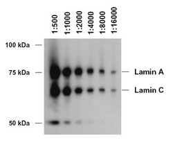
- Western blot analysis of lamin A/C using MAb to Lamin A/C (4C11) (Prod. No. ALX-804-672). Method: HeLa cells were lysed with 1 x GSD boiling buffer. The cell lysate was electrophoretically separated on a 7.5% SDS-PAG and transferred onto a nitrocellulose membrane. One lane contains approximately 7.5µg of whole cell protein. The nitrocellulose membrane was blocked for 1 h with 3% NFDM/PBS-T and then incubated with MAb to Lamin A/C (4C11) at the indicated dilutions (diluted in 0.5% NFDM/PBS-T) for 2 h at RT. The membrane was washed with PBS-T and then incubated for 1 h with HRP coupled anti-mouse antibody (1:5000). The membrane was washed 3 x 10 min with PBS-T and then incubated for 1 min with ECL solution. The signal was detected on a HR-HA 30 X-ray film. Exposure time was 3 min.

- Submitted by
- Enzo Life Sciences (provider)
- Main image
- Experimental details
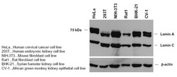
- Western blot analysis of lamin A/C in different species using MAb to Lamin A/C (4C11) (Prod. No. ALX-804-672). Method: GSD-lysates from different species were electrophoretically separated on a 7.5% SDS PAG and blotted on a nitrocellulose membrane. The membrane was blocked with 3% NFDM-PBS/T for 1 hour and incubated withÊMAb to Lamin A/CÊ(4C11)Ê1:1000 in 0.5% NFDM-PBS/T o/N at 4¡C (as loading control anti _-actin, 1:5000). The membrane was washed 3 x 10 min with PBS/T and incubated with anti-mouse HRP, 1:5000 in 0.5% NFDM-PBS/T 1 h at RT. The membrane was washed 3 x 10 min with PBS/T and then incubated for 1 min with ECL solution. The signal was detected on a HR-HA 30 X-ray film. Exposure time was 10 sec.

- Submitted by
- Enzo Life Sciences (provider)
- Main image

- Experimental details
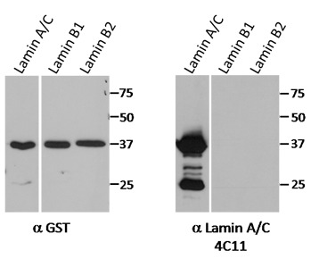
- Lamin A/C, mAb (4C11) (Prod. No. ALX-804-672) does not cross-react with lamin B1 or B2. Method:Western blot analysis of bacterially expressed fusion proteins of GST with the lamin A/C Ig-fold domain (aa 432-544), the lamin B1 Ig-fold domain (aa 434-547), or the lamin B2 Ig-fold domain (aa 466-580). Incubation with a GST-specific antibody (left blot) ensured equal loading. Incubation with the lamin A/C antibody, clone 4C11 (right blot), demonstrates that it does not cross-react with lamin B1 or B2 (even when the film was heavily overexposed as shown in figure). Calculated size of GST-fusion proteins is ~40kDa. The prominent band at ~25kDa represents a degradation product of GST-Lamin A/C-Igfold.
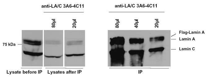
- Submitted by
- Enzo Life Sciences (provider)
- Main image
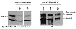
- Experimental details
- Immunoprecipitation of lamin A/C using MAb to Lamin A/C (4C11) (Prod. No. ALX-804-672). Method: For each IP 2mg of whole cell lysate from HeLa cells ectopically expressing FLAG-tagged lamin A were used. The lysates were incubated with the indicated volumes of hybridoma supernatantÊ for 1 hour at 4¡C. Then 30µl of Protein A + G sepharose (1:1 suspension) were added and incubated for 1 hour at 4¡C. Beads were washed, boiled in 30µl 1x GSD boiling buffer for 5 min, and the proteins were loaded on a 10% SDS-PAG and electrophoretically separated (2% of lysate before and after the IP were also loaded). The proteins were blotted on a nitrocellulose membrane. The membrane was blocked with 3% NFDM-PBS/T for 1 hour and then incubated with MAb to Lamin A/C (4C11) (1:1000 in 0.5% NFDM-PBS/T) over night, 4¡C. The membrane was washed 3 x 10 min with PBS/T and then incubated with anti-mouse HRP (1:5000 in 0.5% NFDM-PBS/T) for 1 hour at RT. The membrane was washed 3 x 10 min and then incubated for 1 min with ECL solution. The signal was detected on a HR-HA 30 X-ray film. Exposure time was 5 sec.
- Submitted by
- Enzo Life Sciences (provider)
- Main image

- Experimental details
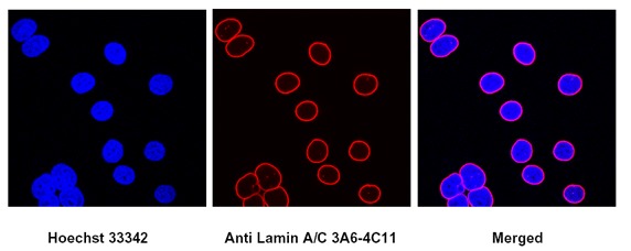
- Immunocytochemistry using MAb to Lamin A/C (4C11) (Prod. No. ALX-804-672). Method: HeLa cells ectopically expressing FLAG-tagged lamin A were grown on coverslips and fixed with ice coldÊmethanol for 10 min at 4¡C, permeabilized with 0.5% Triton-X/PBS for 10 min at RT, blocked with 0.2% gelatine/PBS for 1h and then incubated with MAb to Lamin A/C (4C11) (1:100 in 0.2% gelatine/PBS) for 1 h at RT. Afterwards the cells were incubated with anti-mouse IgG Alexa Fluor 594 antibody (1:500). DNA was counterstained with Hoechst 33342. Pictures were taken with a confocal microscope.