 Explore
 Validate
 Learn
Learn Western blot
Antibody data
- Antibody Data
- Antigen structure
- References [1]
- Comments [0]
- Validations
- Western blot [3]
- Immunocytochemistry [1]
- Immunohistochemistry [1]
Submit
Validation data
Reference
Comment
Report error
- Product number
- PA5-52319 - Provider product page

- Provider
- Invitrogen Antibodies
- Product name
- GBA Polyclonal Antibody
- Antibody type
- Polyclonal
- Antigen
- Recombinant full-length protein
- Description
- The epitope of ABS 006-01-02 is different from BTE 004-09-02 and ABS 006-24-02. The epitope is located on the PAPP-A subunit of the heterotetramer.
- Reactivity
- Human
- Host
- Rabbit
- Isotype
- IgG
- Vial size
- 100 µL
- Concentration
- 0.11 mg/mL
- Storage
- Store at 4°C short term. For long term storage, store at -20°C, avoiding freeze/thaw cycles.
Submitted references In Vivo Remodeling of Altered Autophagy-Lysosomal Pathway by a Phosphopeptide in Lupus.
Wang F, Tasset I, Cuervo AM, Muller S
Cells 2020 Oct 20;9(10)
Cells 2020 Oct 20;9(10)
No comments: Submit comment
Supportive validation
- Submitted by
- Invitrogen Antibodies (provider)
- Main image
- Experimental details
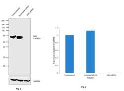
- Knockdown of GBA was achieved by transfecting NIH:OVCAR-3 with GBA specific siRNAs (Silencer® select Product # S501316, S534768). Western Blot analysis (Fig. a) was performed using Whole cell extracts from the GBA knockdown cells (lane 3), non-targeting scrambled siRNA transfected cells (lane 2) and untransfected cells (lane 1). The blot was probed with GBA Polyclonal Antibody (Product # PA5-52319, 1:1000 dilution ) and Goat anti-Rabbit IgG (H+L) Superclonal™ Recombinant Secondary Antibody, HRP (Product # A27036, 1:4000 dilution). Densitometric analysis of this Western Blot is shown in histogram (Fig. b). Loss of signal upon siRNA mediated knock down confirms that antibody is specific to GBA.

- Submitted by
- Invitrogen Antibodies (provider)
- Main image
- Experimental details
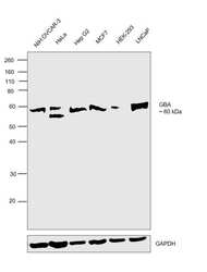
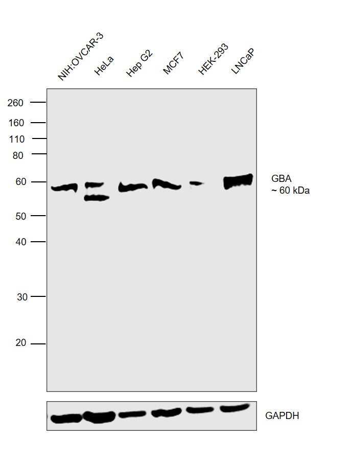
- Western Blot was performed using Anti-GBA Polyclonal Antibody (Product # PA5-52319) and a 60 kDa band corresponding to GBA was observed across the cell lines tested. Whole cell extracts (30 µg lysate) of NIH:OVCAR-3 (Lane 1), HeLa (Lane 2), Hep G2 (Lane 3), MCF7 (Lane 4), HEK-293 (Lane 5) and LNCaP (Lane 6) were electrophoresed using NuPAGE™ 10% Bis-Tris Protein Gel (Product # NP0302BOX). Resolved proteins were then transferred onto a nitrocellulose membrane (Product # IB23001) by iBlot® 2 Dry Blotting System (Product # IB21001). The blot was probed with the primary antibody (0.2 µg/mL) and detected by chemiluminescence with Goat anti-Rabbit IgG (H+L) Superclonal™ Recombinant Secondary Antibody, HRP (Product # A27036, 1:4000 dilution) using the iBright FL 1000 (Product # A32752). Chemiluminescent detection was performed using SuperSignal™ West Dura Extended Duration Substrate (Product # 34076).
- Submitted by
- Invitrogen Antibodies (provider)
- Main image
- Experimental details
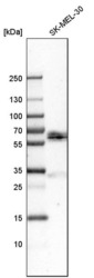
- Western blot analysis of GBA in human cell line SK-MEL-30 using a GBA Polyclonal Antibody (Product # PA5-52319).

Supportive validation
- Submitted by
- Invitrogen Antibodies (provider)
- Main image
- Experimental details
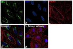
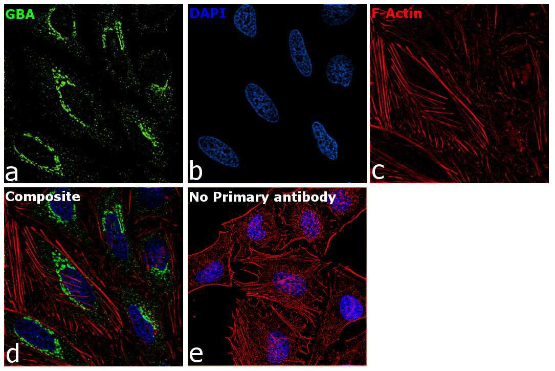
- Immunofluorescence analysis of GBA was performed using 70% confluent log phase HeLa cells. The cells were fixed with 4% paraformaldehyde for 10 minutes, permeabilized with 0.1% Triton™ X-100 for 15 minutes, and blocked with 2% BSA for 45 minutes at room temperature. The cells were labeled with GBA Polyclonal Antibody (Product # PA5-52319) at 1:100 dilution in 0.1% BSA, incubated at 4 degree celsius overnight and then labeled with Donkey anti-Rabbit IgG (H+L) Highly Cross-Adsorbed Secondary Antibody, Alexa Fluor Plus 488 (Product # A32790), (1:2000 dilution), for 45 minutes at room temperature (Panel a: Green). Nuclei (Panel b:Blue) were stained with ProLong™ Diamond Antifade Mountant with DAPI (Product # P36962). F-actin (Panel c: Red) was stained with Rhodamine Phalloidin (Product # R415, 1:300). Panel d represents the merged image showing Lysosomal localization. Panel e represents control cells with no primary antibody to assess background. The images were captured at 60X magnification.
Supportive validation
- Submitted by
- Invitrogen Antibodies (provider)
- Main image
- Experimental details
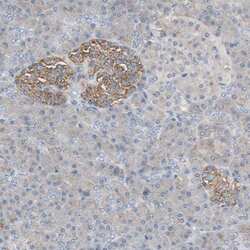
- Immunohistochemical staining of GBA in human pancreas shows strong granular cytoplasmic positivity in islets of Langerhans. Samples were probed using a GBA Polyclonal Antibody (Product # PA5-52319).
