 Explore
 Validate
 Learn
Learn Western blot
Antibody data
- Antibody Data
- Antigen structure
- References [0]
- Comments [0]
- Validations
- Western blot [4]
- Immunocytochemistry [3]
- Immunohistochemistry [2]
Submit
Validation data
Reference
Comment
Report error
- Product number
- PA5-21347 - Provider product page

- Provider
- Invitrogen Antibodies
- Product name
- GBA Polyclonal Antibody
- Antibody type
- Polyclonal
- Antigen
- Recombinant full-length protein
- Description
- Recommended positive controls: 293T, A431, H1299, HeLaS3, HepG2, Molt-4, Raji. Predicted reactivity: Mouse (87%), Rat (91%), Pig (92%), Chimpanzee (100%), Bovine (92%). Store product as a concentrated solution. Centrifuge briefly prior to opening the vial.
- Reactivity
- Human
- Host
- Rabbit
- Isotype
- IgG
- Vial size
- 100 μL
- Concentration
- 1.25 mg/mL
- Storage
- Store at 4°C short term. For long term storage, store at -20°C, avoiding freeze/thaw cycles.
No comments: Submit comment
Supportive validation
- Submitted by
- Invitrogen Antibodies (provider)
- Main image
- Experimental details
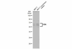
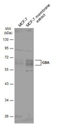
- Western Blot using GBA Polyclonal Antibody (Product # PA5-21347). MCF-7 whole cell and membrane extracts (30 µg) were separated by 10% SDS-PAGE, and the membrane was blotted with GBA Polyclonal Antibody (Product # PA5-21347) diluted at 1:500. The HRP-conjugated anti-rabbit IgG antibody was used to detect the primary antibody.
- Submitted by
- Invitrogen Antibodies (provider)
- Main image

- Experimental details
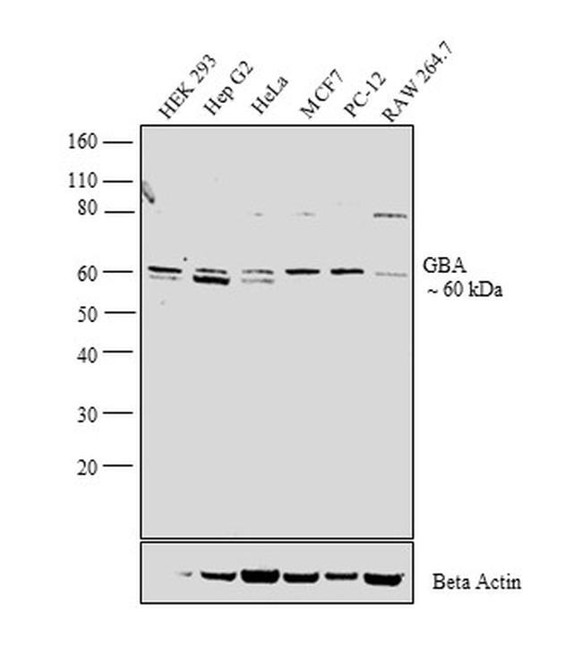
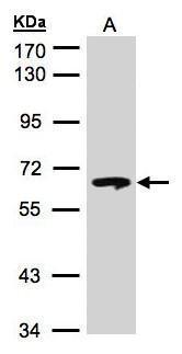
- Western blot analysis was performed on whole cell extracts (30 µg lysate) of HEK 293 (Lane 1), Hep G2 (Lane 2), HeLa (Lane 3), MCF7 (Lane 4), PC-12 (Lane 5) and Raw 264.7 (Lane 6). The blot was probed with Anti- GBA Polyclonal Antibody (Product # PA5-21347, 1:2,000 dilution) and detected by chemiluminescence using Goat anti-Rabbit IgG (Heavy Chain) Superclonal™ Secondary Antibody, HRP conjugate (Product # A27036, 0.25 µg/mL, 1:4,000 dilution). A 60 kDa band corresponding to GBA was observed across all the cell lines tested.
- Submitted by
- Invitrogen Antibodies (provider)
- Main image
- Experimental details
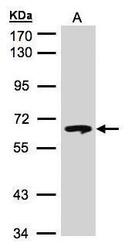
- Western Blot using GBA Polyclonal Antibody (Product # PA5-21347). Sample (30 µg of whole cell lysate). A: Hep G2. 7.5% SDS PAGE. GBA Polyclonal Antibody (Product # PA5-21347) diluted at 1:500. The HRP-conjugated anti-rabbit IgG antibody was used to detect the primary antibody.
- Submitted by
- Invitrogen Antibodies (provider)
- Main image
- Experimental details
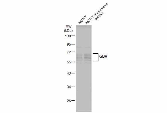
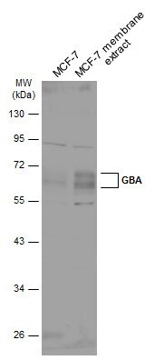
- Western Blot analysis of GBA was performed by separating 30 µg of MCF-7 whole cell and membrane extracts by 10% SDS-PAGE. Proteins were transferred to a membrane and probed with a GBA Polyclonal Antibody (Product # PA5-21347) at a dilution of 1:500 and a HRP-conjugated anti-rabbit IgG secondary antibody.
Supportive validation
- Submitted by
- Invitrogen Antibodies (provider)
- Main image
- Experimental details
- Immunofluorescent analysis of beta-glucosidase in methanol-fixed HeLa cells using a beta-glucosidase polyclonal antibody (Product # PA5-21347) at a 1:100 dilution.

- Submitted by
- Invitrogen Antibodies (provider)
- Main image

- Experimental details
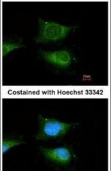
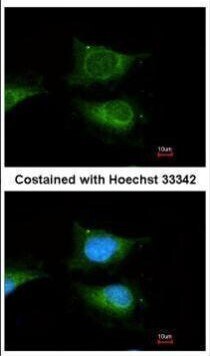
- Immunofluorescence analysis of methanol-fixed HeLa, using GBA antibody (Product # PA5-21347) at 1:100 dilution.

- Submitted by
- Invitrogen Antibodies (provider)
- Main image

- Experimental details
- Immunofluorescence analysis of methanol-fixed HeLa, using GBA antibody (Product # PA5-21347) at 1:100 dilution.
Supportive validation
- Submitted by
- Invitrogen Antibodies (provider)
- Main image
- Experimental details
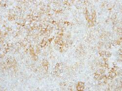
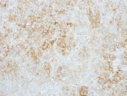
- Immunohistochemical analysis of paraffin-embedded human lung cancer, using GBA (Product # PA5-21347) antibody at 1:100 dilution. Antigen Retrieval: EDTA based buffer, pH 8.0, 15 min.
- Submitted by
- Invitrogen Antibodies (provider)
- Main image
- Experimental details
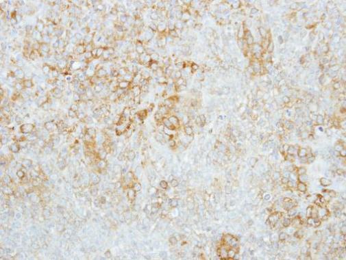
- Immunohistochemical analysis of paraffin-embedded human lung cancer, using GBA (Product # PA5-21347) antibody at 1:100 dilution. Antigen Retrieval: EDTA based buffer, pH 8.0, 15 min.
