Explore
 Validate
 Learn
Learn Western blot
 Immunohistochemistry
Antibody data
- Antibody Data
- Antigen structure
- References [1]
- Comments [0]
- Validations
- Immunohistochemistry [4]
- Other assay [1]
Submit
Validation data
Reference
Comment
Report error
- Product number
- PA5-62944 - Provider product page

- Provider
- Invitrogen Antibodies
- Product name
- KLHL11 Polyclonal Antibody
- Antibody type
- Polyclonal
- Antigen
- Recombinant protein fragment
- Description
- Immunogen sequence: NSDDIDKQYR KEAYRYCAER KRWMLLPPMP QPRCRATACH VRIPYRYLHG TQRYPMPQNL MWQKDRIRQM QEIHRHALNM RRVPSSQIEC Highest antigen sequence identity to the following orthologs: Mouse - 100%, Rat - 100%.
- Reactivity
- Human
- Host
- Rabbit
- Isotype
- IgG
- Vial size
- 100 μL
- Concentration
- 0.1 mg/mL
- Storage
- Store at 4°C short term. For long term storage, store at -20°C, avoiding freeze/thaw cycles.
Submitted references Immunopathogenesis and proposed clinical score for identifying Kelch-like protein-11 encephalitis.
Vogrig A, Péricart S, Pinto AL, Rogemond V, Muñiz-Castrillo S, Picard G, Selton M, Mittelbronn M, Lanoiselée HM, Michenet P, Benaiteau M, Pariente J, Zéphir H, Giordana C, Montaut S, Salhi H, Bachoumas P, Montcuquet A, Letovanec I, Uro-Coste E, Honnorat J
Brain communications 2021;3(3):fcab185
Brain communications 2021;3(3):fcab185
No comments: Submit comment
Supportive validation
- Submitted by
- Invitrogen Antibodies (provider)
- Main image
- Experimental details
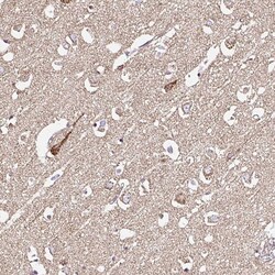
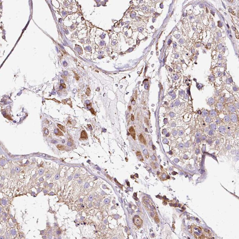
- Immunohistochemical analysis of KLHL11 in human cerebral cortex using KLHL11 Polyclonal Antibody (Product # PA5-62944) shows moderate positivity in neuropil.
- Submitted by
- Invitrogen Antibodies (provider)
- Main image
- Experimental details
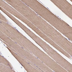
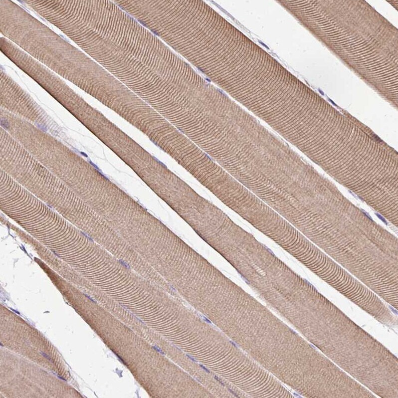
- Immunohistochemical analysis of KLHL11 in human skeletal muscle using KLHL11 Polyclonal Antibody (Product # PA5-62944) shows moderate cytoplasmic positivity in myocytes.
- Submitted by
- Invitrogen Antibodies (provider)
- Main image
- Experimental details
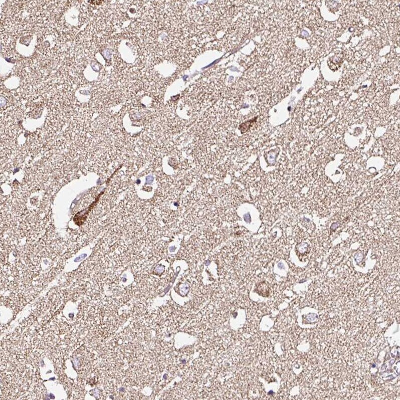
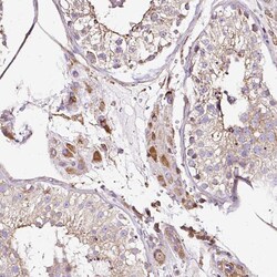
- Immunohistochemical analysis of KLHL11 in human testis using KLHL11 Polyclonal Antibody (Product # PA5-62944) shows moderate cytoplasmic positivity in Leydig cells.
- Submitted by
- Invitrogen Antibodies (provider)
- Main image
- Experimental details
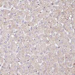
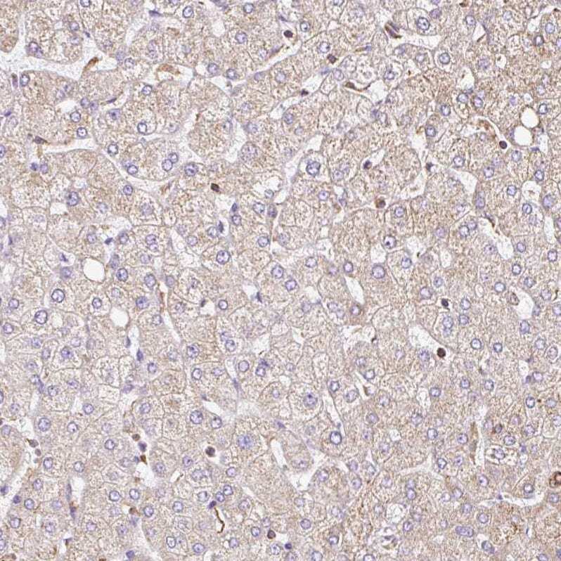
- Immunohistochemical analysis of KLHL11 in human liver using KLHL11 Polyclonal Antibody (Product # PA5-62944) shows very weak positivity in hepatocytes as expected.
Supportive validation
- Submitted by
- Invitrogen Antibodies (provider)
- Main image
- Experimental details
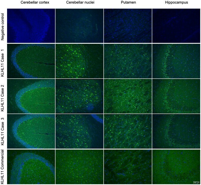
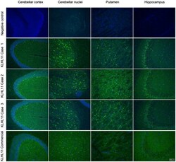
- Figure 1 Immunostaining pattern in patients with KLHL11-Abs. Reactivity of rat brain tissue using a negative control (row 1) and with patient CSF positive for KLHL11-Abs (rows 2, 3 and 4). The CSF of patients showed extensive and diffuse immunostaining involving the cerebellar cortex and nuclei, putamen and hippocampus (in particular, the CA3 region, with a 'comb-like' staining). In the cerebellum and basal ganglia the immunostaining showed a 'leopard-like' appearance. For comparison, immunostaining using a commercial KLHL11-Abs is also shown (row 5), highlighting the involvement of the same regions.