Explore
 Validate
 Learn
Learn Western blot
 Immunocytochemistry
Antibody data
- Antibody Data
- Antigen structure
- References [2]
- Comments [0]
- Validations
- Immunocytochemistry [1]
- Immunohistochemistry [1]
- Chromatin Immunoprecipitation [1]
Submit
Validation data
Reference
Comment
Report error
- Product number
- HPA029273 - Provider product page

- Provider
- Atlas Antibodies
- Proper citation
- Atlas Antibodies Cat#HPA029273, RRID:AB_10603896
- Product name
- Anti-SUPT5H
- Antibody type
- Polyclonal
- Description
- Polyclonal Antibody against Human SUPT5H, Gene description: suppressor of Ty 5 homolog (S. cerevisiae), Alternative Gene Names: FLJ34157, SPT5, SPT5H, Validated applications: WB, IHC, ICC, ChIP, Uniprot ID: O00267, Storage: Store at +4°C for short term storage. Long time storage is recommended at -20°C.
- Reactivity
- Human, Mouse, Rat
- Host
- Rabbit
- Conjugate
- Unconjugated
- Isotype
- IgG
- Vial size
- 100 µl
- Concentration
- 0.1 mg/ml
- Storage
- Store at +4°C for short term storage. Long time storage is recommended at -20°C.
- Handling
- The antibody solution should be gently mixed before use.
Submitted references Suppressor of Ty homolog-5, a novel tumor-specific human telomerase reverse transcriptase promoter-binding protein and activator in colon cancer cells
Proteomic screen reveals Fbw7 as a modulator of the NF-κB pathway
Chen R, Zhu J, Dong Y, He C, Hu X
Oncotarget 2015;6(32):32841-32855
Oncotarget 2015;6(32):32841-32855
Proteomic screen reveals Fbw7 as a modulator of the NF-κB pathway
Arabi A, Ullah K, Branca R, Johansson J, Bandarra D, Haneklaus M, Fu J, Ariës I, Nilsson P, Den Boer M, Pokrovskaja K, Grandér D, Xiao G, Rocha S, Lehtiö J, Sangfelt O
Nature Communications 2012;3(1)
Nature Communications 2012;3(1)
No comments: Submit comment
Supportive validation
- Submitted by
- Atlas Antibodies (provider)
- Main image
- Experimental details
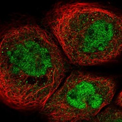
- Immunofluorescent staining of human cell line A-431 shows localization to nucleoplasm.
- Sample type
- Human

Supportive validation
- Submitted by
- Atlas Antibodies (provider)
- Enhanced method
- Orthogonal validation
- Main image

- Experimental details
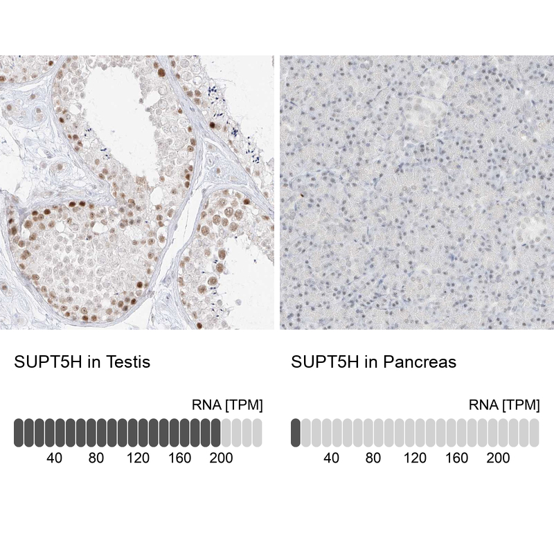
- Immunohistochemistry analysis in human testis and pancreas tissues using Anti-SUPT5H antibody. Corresponding SUPT5H RNA-seq data are presented for the same tissues.
- Sample type
- Human
- Protocol
- Protocol
Supportive validation
- Submitted by
- Atlas Antibodies (provider)
- Main image
- Experimental details
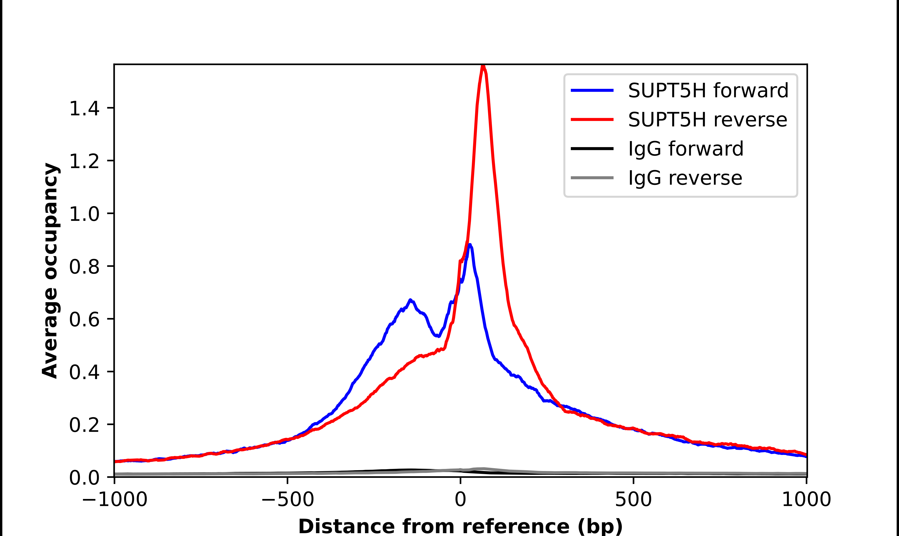
- ChIP-Exo-Seq composite graph for Anti-SUPT5H (HPA029273, Lot 000012280) tested in K562 cells. Strand-specific reads (blue: forward, red: reverse) and IgG controls (black: forward, grey: reverse) are plotted against the distance from a composite set of reference binding sites. The antibody exhibits robust target enrichment compared to a non-specific IgG control and precisely reveals its structural organization around the binding site. Data generated by Prof. B. F. Pugh´s Lab at Cornell University.