 Explore
 Validate
 Learn
Learn Western blot
Antibody data
- Antibody Data
- Antigen structure
- References [0]
- Comments [0]
- Validations
- Western blot [3]
- Immunohistochemistry [3]
Submit
Validation data
Reference
Comment
Report error
- Product number
- PA5-40709 - Provider product page

- Provider
- Invitrogen Antibodies
- Product name
- SPT5 Polyclonal Antibody
- Antibody type
- Polyclonal
- Antigen
- Synthetic peptide
- Description
- Peptide sequence: PYAAPSPQGS YQPSPSPQSY HQVAPSPAGY QNTHSPASYH PTPSPMAYQA Sequence homology: Cow: 100%; Dog: 100%; Guinea Pig: 100%; Horse: 100%; Human: 100%; Rabbit: 100%; Zebrafish: 92%
- Reactivity
- Human
- Host
- Rabbit
- Isotype
- IgG
- Vial size
- 100 µL
- Concentration
- 0.5 mg/mL
- Storage
- -20° C, Avoid Freeze/Thaw Cycles
No comments: Submit comment
Supportive validation
- Submitted by
- Invitrogen Antibodies (provider)
- Main image
- Experimental details
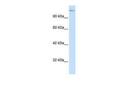
- Western blot analysis of human HepG2 cell lysate using an anti-SUPT5H polyclonal antibody (Product # PA5-40709).

- Submitted by
- Invitrogen Antibodies (provider)
- Main image
- Experimental details
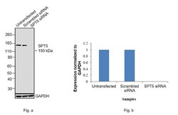
- Knockdown of SPT5 was achieved by transfecting HeLa with SPT5 specific siRNAs (Silencer® select Product # s13631, s13632). Western blot analysis (Fig. a) was performed using whole cell extracts from the SPT5 knockdown cells (lane 3), non-specific scrambled siRNA transfected cells (lane 2) and untransfected cells (lane 1). The blot was probed with SPT5 Polyclonal Antibody (Product # PA5-40709, 0.5 ug/ml) and Goat anti-Rabbit IgG (H+L) Superclonal™ Secondary Antibody, HRP (Product # A27036, 0.25µg/ml, 1:4000 dilution). Densitometric analysis of this western blot is shown in histogram (Fig. b). Decrease in signal upon siRNA mediated knock down confirms that antibody is specific to SPT5.

- Submitted by
- Invitrogen Antibodies (provider)
- Main image
- Experimental details
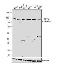
- Western blot was performed using Anti-SPT5 Polyclonal Antibody (Product # PA5-40709) and a 150 kDa band corresponding to SPT5 was observed across all the cell lines tested. Modified whole cell extracts (1% SDS) (30 µg lysate) of HeLa (Lane 1), Hep G2 (Lane 2), A-431 (Lane 3), A549 (Lane 4), HT-29 (Lane 5) and K-562 (Lane 6) were electrophoresed using NuPAGE™ 4-12% Bis-Tris Protein Gel (Product # NP0322BOX). Resolved proteins were then transferred onto a nitrocellulose membrane (Product # IB23001) by XCell SureLock™ Mini-Cell and XCell II™ Blot Module (Product # EI0002). The blot was probed with the primary antibody (0.5 ug/ml) and detected by chemiluminescence with Goat Anti-Rabbit IgG Secondary Antibody, HRP conjugate (Product # A27036, 1:4000 dilution) using the iBright FL 1000 (Product # A32752). Chemiluminescent detection was performed using Novex® ECL Chemiluminescent Substrate Reagent Kit (Product # WP20005).

Supportive validation
- Submitted by
- Invitrogen Antibodies (provider)
- Main image

- Experimental details
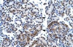
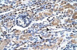
- Immunohistochemistry (paraffin-embedded) analysis of human alveolar tissue using an anti-SUPT5H polyclonal antibody (Product # PA5-40709).

- Submitted by
- Invitrogen Antibodies (provider)
- Main image
- Experimental details
- Immunohistochemistry (paraffin-embedded) analysis of human pancreas tissue using an anti-SUPT5H polyclonal antibody (Product # PA5-40709).

- Submitted by
- Invitrogen Antibodies (provider)
- Main image
- Experimental details
- Immunohistochemistry (paraffin-embedded) analysis of human kidney tissue using an anti-SUPT5H polyclonal antibody (Product # PA5-40709).
