 Explore
 Validate
 Learn
Learn Western blot
Antibody data
- Antibody Data
- Antigen structure
- References [1]
- Comments [0]
- Validations
- Western blot [3]
- Immunohistochemistry [1]
Submit
Validation data
Reference
Comment
Report error
- Product number
- AF4584 - Provider product page

- Provider
- R&D Systems
- Product name
- Human/Mouse S100A6 Antibody
- Antibody type
- Polyclonal
- Description
- Antigen Affinity-purified. Detects human and mouse S100A6 in direct ELISAs and Western blots. In direct ELISAs, less than 2% cross-reactivity with recombinant mouse S100A1 and recombinant mouse S100A4 is observed.
- Reactivity
- Human, Mouse
- Host
- Sheep
- Conjugate
- Unconjugated
- Antigen sequence
P14069- Isotype
- IgG
- Vial size
- 100 ug
- Concentration
- LYOPH
- Storage
- Use a manual defrost freezer and avoid repeated freeze-thaw cycles. 12 months from date of receipt, -20 to -70 °C as supplied. 1 month, 2 to 8 °C under sterile conditions after reconstitution. 6 months, -20 to -70 °C under sterile conditions after reconstitution.
Submitted references MMP-8 deficiency increases TLR/RAGE ligands S100A8 and S100A9 and exacerbates lung inflammation during endotoxemia.
González-López A, Aguirre A, López-Alonso I, Amado L, Astudillo A, Fernández-García MS, Suárez MF, Batalla-Solís E, Colado E, Albaiceta GM
PloS one 2012;7(6):e39940
PloS one 2012;7(6):e39940
No comments: Submit comment
Supportive validation
- Submitted by
- R&D Systems (provider)
- Main image
- Experimental details
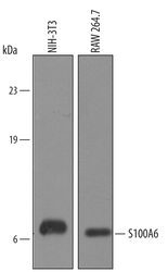
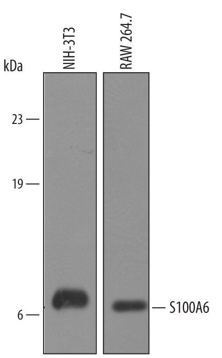
- Detection of Mouse S100A6 by Western Blot. Western blot shows lysates of RAW 264.7 mouse monocyte/macrophage cell line and NIH-3T3 mouse embryonic fibroblast cell line. PVDF membrane was probed with 1 µg/mL of Sheep Anti-Human/ Mouse S100A6 Antigen Affinity-purified Polyclonal Antibody (Catalog # AF4584) followed by HRP-conjugated Anti-Sheep IgG Secondary Antibody (Catalog # HAF016). A specific band was detected for S100A6 at approximately 10 kDa (as indicated). This experiment was conducted under reducing conditions and using Immunoblot Buffer Group 8.
- Submitted by
- R&D Systems (provider)
- Main image
- Experimental details
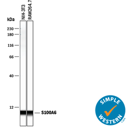
- Detection of Mouse S100A6 by Simple WesternTM. Simple Western lane view shows lysates of NIH-3T3 mouse embryonic fibroblast cell line and RAW 264.7 mouse monocyte/macrophage cell line, loaded at 0.2 mg/mL. A specific band was detected for S100A6 at approximately 7 kDa (as indicated) using 10 µg/mL of Sheep Anti-Human/Mouse S100A6 Antigen Affinity-purified Polyclonal Antibody (Catalog # AF4584) followed by 1:50 dilution of HRP-conjugated Anti-Sheep IgG Secondary Antibody (Catalog # HAF016). This experiment was conducted under reducing conditions and using the 12-230 kDa separation system.

- Submitted by
- R&D Systems (provider)
- Main image

- Experimental details
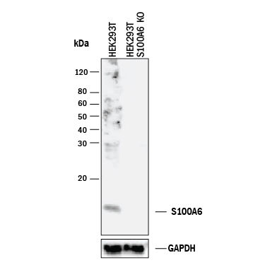
- Western Blot Shows Human S100A6 Specificity by Using Knockout Cell Line. Western blot shows lysates of HEK293T human embryonic kidney parental cell line and S100A6 knockout HEK293T cell line (KO). PVDF membrane was probed with 1 µg/mL of Sheep Anti-Human/Mouse S100A6 Antigen Affinity-purified Polyclonal Antibody (Catalog # AF4584) followed by HRP-conjugated Anti-Sheep IgG Secondary Antibody (Catalog # HAF016). A specific band was detected for S100A6 at approximately 10 kDa (as indicated) in the parental HEK293T cell line, but is not detectable in knockout HEK293T cell line. GAPDH (Catalog # AF5718) is shown as a loading control. This experiment was conducted under reducing conditions and using Immunoblot Buffer Group 1.
Supportive validation
- Submitted by
- R&D Systems (provider)
- Main image
- Experimental details
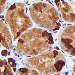
- S100A6 in Human Kidney. S100A6 was detected in immersion fixed paraffin-embedded sections of human kidney using Sheep Anti-Human/Mouse S100A6 Antigen Affinity-purified Polyclonal Antibody (Catalog # AF4584) at 3 µg/mL overnight at 4 °C. Before incubation with the primary antibody, tissue was subjected to heat-induced epitope retrieval using Antigen Retrieval Reagent-Basic (Catalog # CTS013). Tissue was stained using the Anti-Sheep HRP-DAB Cell & Tissue Staining Kit (brown; Catalog # CTS019) and counterstained with hematoxylin (blue). Specific staining was localized to epithelial cells in convoluted tubules. View our protocol for Chromogenic IHC Staining of Paraffin-embedded Tissue Sections.
