Explore
 Validate
 Learn
Learn Western blot
Antibody data
- Antibody Data
- Antigen structure
- References [2]
- Comments [0]
- Validations
- Western blot [1]
- Immunocytochemistry [2]
- Immunohistochemistry [3]
- Other assay [1]
Submit
Validation data
Reference
Comment
Report error
- Product number
- PA1-32229 - Provider product page

- Provider
- Invitrogen Antibodies
- Product name
- TACR1 Polyclonal Antibody
- Antibody type
- Polyclonal
- Antigen
- Synthetic peptide
- Description
- Store product as a concentrated solution. Centrifuge briefly prior to opening the vial.
- Reactivity
- Human, Mouse, Rat, Canine, Guinea Pig
- Host
- Rabbit
- Isotype
- IgG
- Vial size
- 100 μL
- Concentration
- Conc. Not Determined
- Storage
- Store at 4°C short term. For long term storage, store at -20°C, avoiding freeze/thaw cycles.
Submitted references Activation of neuroimmune pathways increases therapeutic effects of radiotherapy on poorly differentiated breast carcinoma.
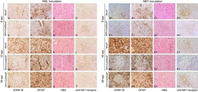
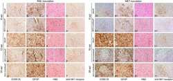
Identification of clinical target areas in the brainstem of prion-infected mice.
Erin N, Korcum AF, Tanrıöver G, Kale Ş, Demir N, Köksoy S
Brain, behavior, and immunity 2015 Aug;48:174-85
Brain, behavior, and immunity 2015 Aug;48:174-85
Identification of clinical target areas in the brainstem of prion-infected mice.
Mirabile I, Jat PS, Brandner S, Collinge J
Neuropathology and applied neurobiology 2015 Aug;41(5):613-30
Neuropathology and applied neurobiology 2015 Aug;41(5):613-30
No comments: Submit comment
Supportive validation
- Submitted by
- Invitrogen Antibodies (provider)
- Main image
- Experimental details
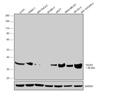
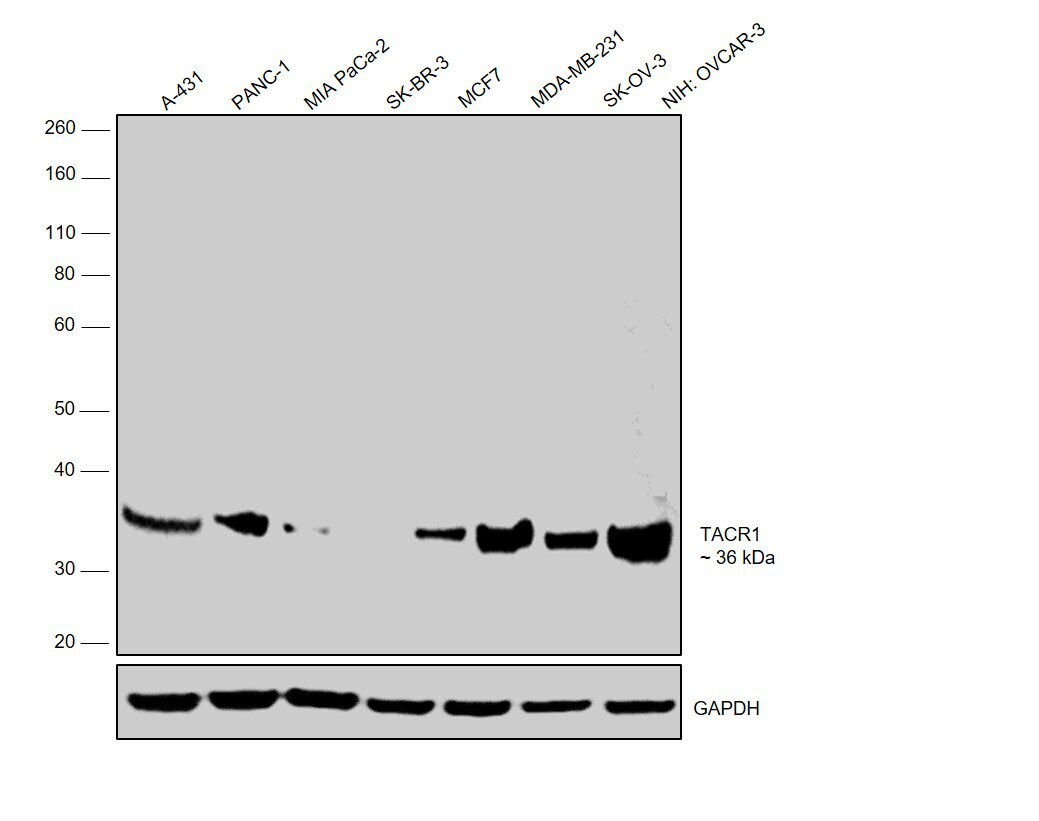
- Western blot was performed using Anti-TACR1 Polyclonal Antibody (Product # PA1-32229) and 36 kDa bands corresponding to TACR1 were observed across cell lines tested except in MIA-PaCa-2 and SK-BR-3, which are reported to be low expressing for TACR1. Whole cell extracts (30 µg lysate) of A-431 (Lane 1), PANC-1 (Lane 2), MIA-PaCa-2 (Lane 3), SK-BR-3 (Lane 4), MCF7 (Lane 5), MDA-MB-231 (Lane 6), SK-OV-3 (Lane 7) and NIH: OVCAR-3 (Lane 8) were electrophoresed using Novex® NuPAGE® 12 % Bis-Tris gel (Product # NP0342BOX). Resolved proteins were then transferred onto a nitrocellulose membrane (Product # IB23001) by iBlot® 2 Dry Blotting System (Product # IB21001). The blot was probed with the primary antibody (1:1000 dilution) and detected by chemiluminescence with Goat anti-Rabbit IgG (Heavy Chain), Superclonal™ Recombinant Secondary Antibody, HRP (Product # A27036, 1:4000 dilution) using the iBright FL 1000 (Product # A32752). Chemiluminescent detection was performed using Novex® ECL Chemiluminescent Substrate Reagent Kit (Product # WP20005)..
Supportive validation
- Submitted by
- Invitrogen Antibodies (provider)
- Main image

- Experimental details
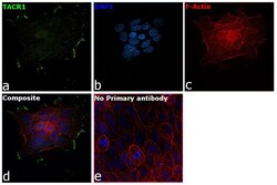
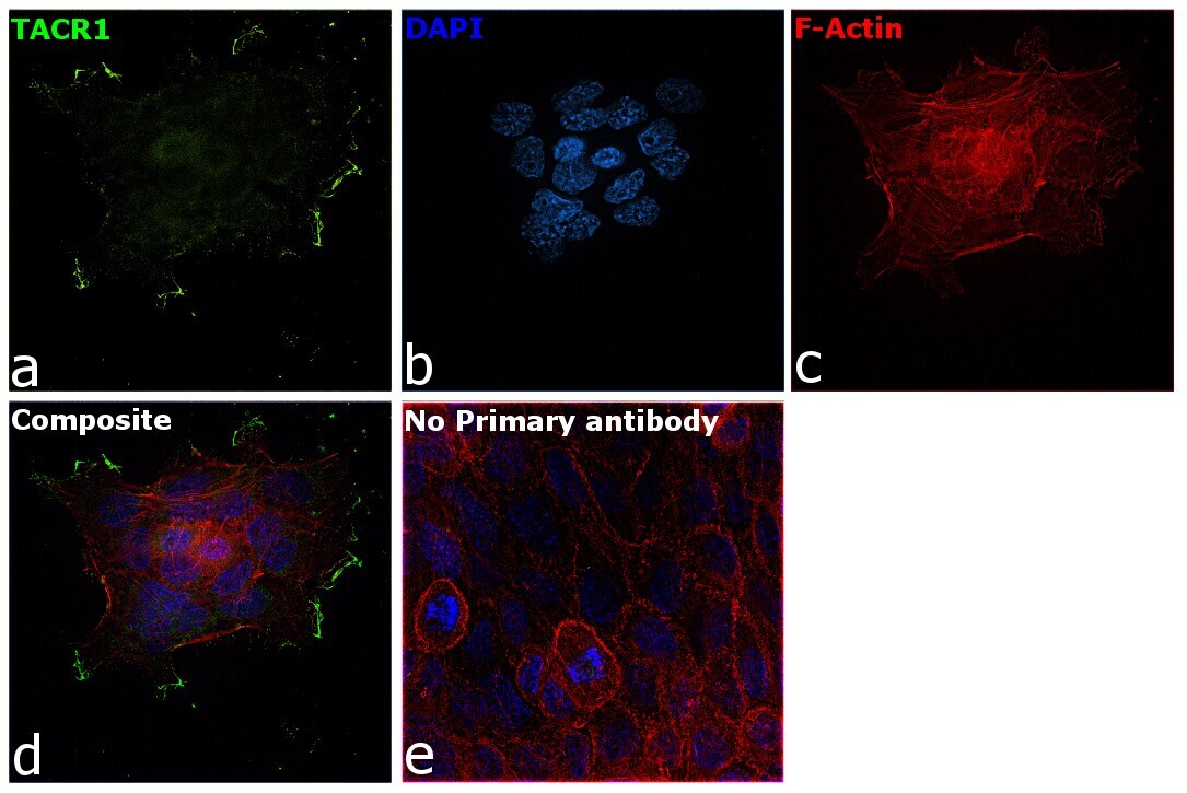
- Immunofluorescence analysis of TACR1 was performed using 70% confluent log phase A-431 cells. The cells were fixed with 4% paraformaldehyde for 10 minutes, permeabilized with 0.1% Triton™ X-100 for 15 minutes, and blocked with 2% BSA for 1 hour at room temperature. The cells were labeled with TACR1 Polyclonal Antibody (Product PA1-32229) at 1:100 dilution in 0.1% BSA, incubated at 4 degree celsius overnight and then with Goat anti-Rabbit IgG (H+L), Superclonal™ Recombinant Secondary Antibody, Alexa Fluor 488 (Product # A27034, 1:2000 dilution) for 45 minutes at room temperature (Panel a: Green). Nuclei (Panel b: Blue) were stained with SlowFade® Gold Antifade Mountant with DAPI (Product # S36938). F-actin (Panel c: Red) was stained with Rhodamine Phalloidin (Product # R415, 1:300). Panel d represents the merged image showing cytoplasmic and membrane localization of TACR1. Panel e represents control cells with no primary antibody to assess background. The images were captured at 60X magnification.

- Submitted by
- Invitrogen Antibodies (provider)
- Main image
- Experimental details
- Immunofluorescence analysis of TACR1 was performed using 70% confluent log phase A-431 cells. The cells were fixed with 4% paraformaldehyde for 10 minutes, permeabilized with 0.1% Triton™ X-100 for 15 minutes, and blocked with 2% BSA for 1 hour at room temperature. The cells were labeled with TACR1 Polyclonal Antibody (Product PA1-32229) at 1:100 dilution in 0.1% BSA, incubated at 4 degree celsius overnight and then with Goat anti-Rabbit IgG (Heavy Chain), Superclonal™ Recombinant Secondary Antibody, Alexa Fluor 488 (Product # A27034, 1:2000 dilution) for 45 minutes at room temperature (Panel a: Green). Nuclei (Panel b: Blue) were stained with SlowFade® Gold Antifade Mountant with DAPI (Product # S36938). F-actin (Panel c: Red) was stained with Rhodamine Phalloidin (Product # R415, 1:300). Panel d represents the merged image showing cytoplasmic and membrane localization of TACR1. Panel e represents control cells with no primary antibody to assess background. The images were captured at 60X magnification.
Supportive validation
- Submitted by
- Invitrogen Antibodies (provider)
- Main image
- Experimental details
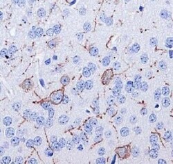
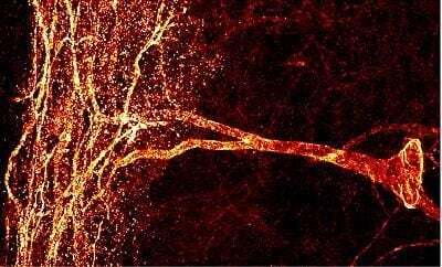
- Immunohistochemistry (Paraffin) analysis of TACR1 in mouse brain (selected cells, axon and dendrites) tissue using TACR1 Polyclonal Antibody (Product # PA1-32229).
- Submitted by
- Invitrogen Antibodies (provider)
- Main image

- Experimental details
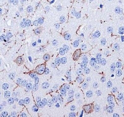
- Immunohistochemistry (Paraffin) analysis of TACR1 in rat spinal cord tissue using TACR1 Polyclonal Antibody (Product # PA1-32229).

- Submitted by
- Invitrogen Antibodies (provider)
- Main image
- Experimental details
- Immunohistochemistry (Paraffin) analysis of TACR1 in rat spinal cord tissue using TACR1 Polyclonal Antibody (Product # PA1-32229).
Supportive validation
- Submitted by
- Invitrogen Antibodies (provider)
- Main image
- Experimental details
- NULL