 Explore
 Validate
 Learn
LearnPA1-18277
antibody from Invitrogen Antibodies
Targeting: PRDX6
1-Cys, aiPLA2, AOP2, KIAA0106, MGC46173, NSGPx, p29, PRX
 Western blot
 ELISA
ELISAAntibody data
- Antibody Data
- Antigen structure
- References [0]
- Comments [0]
- Validations
- Western blot [2]
Submit
Validation data
Reference
Comment
Report error
- Product number
- PA1-18277 - Provider product page

- Provider
- Invitrogen Antibodies
- Product name
- PRDX6 Polyclonal Antibody
- Antibody type
- Polyclonal
- Antigen
- Recombinant full-length protein
- Description
- Reconstitute in 100 µL of sterile water. Centrifuge to remove any insoluble material. After reconstitution keep aliquots at -20 °C for a higher stability, and at 4 °C with an appropriate antibacterial agent. Avoid repetitive freeze/thaw cycles. Glycerol (1:1) may be added for an additional stability.
- Reactivity
- Human, Mouse, Rat
- Host
- Rabbit
- Isotype
- IgG
- Vial size
- 100 µL
- Concentration
- Conc. Not Determined
- Storage
- -20° C, Avoid Freeze/Thaw Cycles
No comments: Submit comment
Supportive validation
- Submitted by
- Invitrogen Antibodies (provider)
- Main image
- Experimental details
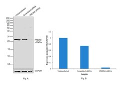
- Knockdown of PRDX6 was achieved by transfecting MCF7 with PRDX6 specific siRNAs (Silencer® select Product # S18428, S18429). Western blot analysis (Fig. a) was performed using Whole cell extracts from the PRDX6 knockdown cells (lane 3), non-targeting scrambled siRNA transfected cells (lane 2) and untransfected cells (lane 1). The blot was probed with PRDX6 Polyclonal Antibody (Product # PA1-18277, 1:1000) and Goat anti-Rabbit IgG (H+L) Superclonal™ Recombinant Secondary Antibody, HRP (Product # A27036, 1:4000). Densitometric analysis of this western blot is shown in histogram (Fig. b). Decrease in signal upon siRNA mediated knock down confirms that antibody is specific to PRDX6.

- Submitted by
- Invitrogen Antibodies (provider)
- Main image

- Experimental details
- Western blot was performed using Anti-PRDX6 Polyclonal Antibody(Product # PA1-18277) and a 25kDa band corresponding to PRDX6 was observed across all the tested cell lines. Whole cell extracts (30 µg lysate) of MCF7 (Lane 1), HeLa (Lane 2), NTERA-2 cl.D1 (Lane 3), Caco-2 (Lane 4), Hep G2 (Lane 5), LNCaP (Lane 6), NIH/3T3 (Lane 7), PC-12 (Lane 8) were electrophoresed using NuPAGE™ 12% Bis-Tris Protein Gel (Product # NP0342BOX). Resolved proteins were then transferred onto a Nitrocellulose membrane (Product # IB23001) by iBlot® 2 Dry Blotting System (Product # IB21001). The blot was probed with the primary antibody (1:2000) and detected by chemiluminescence with Goat anti-Rabbit IgG (H+L) Superclonal™ Recombinant Secondary Antibody, HRP (Product # A27036, 1:4000) using the iBright FL 1000 (Product # A32752). Chemiluminescent detection was performed using Novex® ECL Chemiluminescent Substrate Reagent Kit (Product # WP20005).
