 Explore
 Validate
 Learn
Learn Western blot
 ELISA
ELISAAntibody data
- Antibody Data
- Antigen structure
- References [5]
- Comments [0]
- Validations
- Western blot [2]
- Immunocytochemistry [2]
- Immunohistochemistry [3]
Submit
Validation data
Reference
Comment
Report error
- Product number
- MA1-413 - Provider product page

- Provider
- Invitrogen Antibodies
- Product name
- Phospho-Progesterone Receptor (Ser190) Monoclonal Antibody (1154)
- Antibody type
- Monoclonal
- Antigen
- Synthetic peptide
- Description
- MA1-413 detects phospho-progesterone receptor (pSer190) from human samples. MA1-413 has been successfully used in Western blot, immunohistochemistry, ELISA, immunofluorescence, and immunoprecipitation procedures. By Western blot, this antibody detects ~81 and ~116 kDa proteins representing phospho-progesterone receptor (pSer190) A and B from T47D cell and human breast carcinoma extracts. Immunohistochemical staining of phospho-progesterone receptor (pSer190) (paraffin samples) can be performed using MA1-413. The MA1-413 immunogen is a synthetic peptide corresponding to residues V(184) L P R G L S(p) P A R Q L L(196) of human Progesterone Receptor.
- Reactivity
- Human
- Host
- Mouse
- Isotype
- IgG
- Antibody clone number
- 1154
- Vial size
- 100 µg
- Concentration
- 1 mg/mL
- Storage
- -20° C, Avoid Freeze/Thaw Cycles
Submitted references Classical membrane progesterone receptors in murine mammary carcinomas: agonistic effects of progestins and RU-486 mediating rapid non-genomic effects.
Mutational analysis of progesterone receptor functional domains in stable cell lines delineates sets of genes regulated by different mechanisms.
Carcinoma-associated fibroblasts activate progesterone receptors and induce hormone independent mammary tumor growth: A role for the FGF-2/FGFR-2 axis.
Identification of a novel mitotic phosphorylation motif associated with protein localization to the mitotic apparatus.
Differential hormone-dependent phosphorylation of progesterone receptor A and B forms revealed by a phosphoserine site-specific monoclonal antibody.
Bottino MC, Cerliani JP, Rojas P, Giulianelli S, Soldati R, Mondillo C, Gorostiaga MA, Pignataro OP, Calvo JC, Gutkind JS, Panomwat Amornphimoltham, Molinolo AA, Lüthy IA, Lanari C
Breast cancer research and treatment 2011 Apr;126(3):621-36
Breast cancer research and treatment 2011 Apr;126(3):621-36
Mutational analysis of progesterone receptor functional domains in stable cell lines delineates sets of genes regulated by different mechanisms.
Quiles I, Millán-Ariño L, Subtil-Rodríguez A, Miñana B, Spinedi N, Ballaré C, Beato M, Jordan A
Molecular endocrinology (Baltimore, Md.) 2009 Jun;23(6):809-26
Molecular endocrinology (Baltimore, Md.) 2009 Jun;23(6):809-26
Carcinoma-associated fibroblasts activate progesterone receptors and induce hormone independent mammary tumor growth: A role for the FGF-2/FGFR-2 axis.
Giulianelli S, Cerliani JP, Lamb CA, Fabris VT, Bottino MC, Gorostiaga MA, Novaro V, Góngora A, Baldi A, Molinolo A, Lanari C
International journal of cancer 2008 Dec 1;123(11):2518-31
International journal of cancer 2008 Dec 1;123(11):2518-31
Identification of a novel mitotic phosphorylation motif associated with protein localization to the mitotic apparatus.
Yang F, Camp DG 2nd, Gritsenko MA, Luo Q, Kelly RT, Clauss TR, Brinkley WR, Smith RD, Stenoien DL
Journal of cell science 2007 Nov 15;120(Pt 22):4060-70
Journal of cell science 2007 Nov 15;120(Pt 22):4060-70
Differential hormone-dependent phosphorylation of progesterone receptor A and B forms revealed by a phosphoserine site-specific monoclonal antibody.
Clemm DL, Sherman L, Boonyaratanakornkit V, Schrader WT, Weigel NL, Edwards DP
Molecular endocrinology (Baltimore, Md.) 2000 Jan;14(1):52-65
Molecular endocrinology (Baltimore, Md.) 2000 Jan;14(1):52-65
No comments: Submit comment
Supportive validation
- Submitted by
- Invitrogen Antibodies (provider)
- Main image

- Experimental details
- Western blot analysis of Phosphorylated Progesterone Receptor (upper panel) and total Progesterone Receptor (lower panel) was performed by loading 20 µg of nuclear (N) or cytoplasmic (C) T47D cell lysates, untreated (-) or stimulated (+) with 100 nm promegestone (R5020) for 1 hour and 10 µL PageRuler Plus Prestained Protein Ladder (Product # 26619) per well onto a 4-20% Tris-Glycine polyacrylamide gel. Proteins were transferred to a nitrocellulose membrane using the G2 Fast Blotter (Product # 62288) and blocked with 5% Milk/TBST for at least 1 hour at room temperature. Phosphorylated Progesterone Receptor was detected using a Phospho-Progesterone Receptor (Ser190) mouse monoclonal antibody (upper panel, Product # MA1-413) at a concentration of 4 µg/mL and Total Progesterone Receptor was detected using a Progesterone Receptor mouse monoclonal antibody (lower panel, Product # MA1-12626) at a concentration of 1 µg/mL in blocking buffer overnight at 4°C on a rocking platform, followed by a Superclonal goat anti-Mouse IgG-HRP secondary antibody (Product # A28177) at a dilution of 1:2,000 for at least 1 hour at room temperature. Chemiluminescent detection was performed using SuperSignal West Pico (Product # 34078) and the myECL Imager (Product # 62236).

Supportive validation
- Submitted by
- Invitrogen Antibodies (provider)
- Main image
- Experimental details
- Immunofluorescent analysis of Phospho-Progesterone Receptor pSer190 in untreated T47D cells (left panel) or stimulated cells with 100 nM of Progesterone (right panel). Formalin-fixed cells were permeabilized with 0.1% Triton X-100 in TBS for 5-10 minutes at room temperature and blocked with 3% BSA-PBS for 30 minutes at room temperature. Cells were probed with a Phospho-Progesterone Receptor pSer190 Monclonal Antibody (1154) (Product # MA1-413) at a dilution of 1:50 and incubated overnight in a humidified chamber. Cells were washed with PBST and incubated with a DyLight-conjugated secondary antibody for 45 minutes at room temperature in the dark. F-actin (red) was stained with a fluorescent phalloidin and nuclei (blue) were stained with DAPI. Images were taken at a 60X magnification.

- Submitted by
- Invitrogen Antibodies (provider)
- Main image

- Experimental details
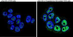
- Immunofluorescent analysis of Phospho-Progesterone Receptor pSer190 (green) showing staining in T47D cells (right) compared to a negative control without primary antibody (left). Formalin-fixed cells were permeabilized with 0.1% Triton X-100 in TBS for 5-10 minutes and blocked with 3% BSA-PBS for 30 minutes at room temperature. Cells were probed with a Phospho-Progesterone Receptor pSer190 monoclonal antibody (Product # MA1-413) in 3% BSA-PBS at a dilution of 1:50 and incubated overnight at 4ºC in a humidified chamber. Cells were washed with PBST and incubated with a DyLight-conjugated secondary antibody in PBS at room temperature in the dark. Nuclei (blue) were stained with Hoechst or DAPI. Images were taken at a magnification of 60x.

Supportive validation
- Submitted by
- Invitrogen Antibodies (provider)
- Main image

- Experimental details
- Immunohistochemistry was performed on cancer biopsies of deparaffinized Human breast carcinoma tissue. To expose target proteins, heat induced antigen retrieval was performed using 10mM sodium citrate (pH6.0) buffer, microwaved for 8-15 minutes. Following antigen retrieval tissues were blocked in 3% BSA-PBS for 30 minutes at room temperature. Tissues were then probed at a dilution of 1:20 with a mouse monoclonal antibody recognizing Phospho-Progesterone Receptor pSer190 (Product # MA1-413) or without primary antibody (negative control) overnight at 4°C in a humidified chamber. Tissues were washed extensively with PBST and endogenous peroxidase activity was quenched with a peroxidase suppressor. Detection was performed using a biotin-conjugated secondary antibody and SA-HRP, followed by colorimetric detection using DAB. Tissues were counterstained with hematoxylin and prepped for mounting.

- Submitted by
- Invitrogen Antibodies (provider)
- Main image

- Experimental details
- Immunohistochemistry was performed on biopsies of deparaffinized Human tonsil tissue. To expose target proteins, heat induced antigen retrieval was performed using 10mM sodium citrate (pH6.0) buffer, microwaved for 8-15 minutes. Following antigen retrieval tissues were blocked in 3% BSA-PBS for 30 minutes at room temperature. Tissues were then probed at a dilution of 1:20 with a mouse monoclonal antibody recognizing Phospho-Progesterone Receptor pSer190 (Product # MA1-413) or without primary antibody (negative control) overnight at 4°C in a humidified chamber. Tissues were washed extensively with PBST and endogenous peroxidase activity was quenched with a peroxidase suppressor. Detection was performed using a biotin-conjugated secondary antibody and SA-HRP, followed by colorimetric detection using DAB. Tissues were counterstained with hematoxylin and prepped for mounting.

- Submitted by
- Invitrogen Antibodies (provider)
- Main image

- Experimental details
- Immunohistochemistry was performed on biopsies of deparaffinized Human uterus tissue. To expose target proteins, heat induced antigen retrieval was performed using 10mM sodium citrate (pH6.0) buffer, microwaved for 8-15 minutes. Following antigen retrieval tissues were blocked in 3% BSA-PBS for 30 minutes at room temperature. Tissues were then probed at a dilution of 1:200 with a mouse monoclonal antibody recognizing Phospho-Progesterone Receptor pSer190 (Product # MA1-413) or without primary antibody (negative control) overnight at 4°C in a humidified chamber. Tissues were washed extensively with PBST and endogenous peroxidase activity was quenched with a peroxidase suppressor. Detection was performed using a biotin-conjugated secondary antibody and SA-HRP, followed by colorimetric detection using DAB. Tissues were counterstained with hematoxylin and prepped for mounting.
