Explore
 Validate
 Learn
Learn Western blot
Antibody data
- Antibody Data
- Antigen structure
- References [11]
- Comments [0]
- Validations
- Western blot [1]
- Immunocytochemistry [3]
- Flow cytometry [1]
Submit
Validation data
Reference
Comment
Report error
- Product number
- MA1-414 - Provider product page

- Provider
- Invitrogen Antibodies
- Product name
- Phospho-Progesterone Receptor (Ser294) Monoclonal Antibody (608)
- Antibody type
- Monoclonal
- Antigen
- Synthetic peptide
- Description
- MA1-414 detects phospho-progesterone receptor (pSer294) from human samples.
- Antibody clone number
- 608
- Concentration
- 1 mg/mL
Submitted references A Common Docking Domain in Progesterone Receptor-B links DUSP6 and CK2 signaling to proliferative transcriptional programs in breast cancer cells.
Classical membrane progesterone receptors in murine mammary carcinomas: agonistic effects of progestins and RU-486 mediating rapid non-genomic effects.
Immunoneutralization of inhibin in cycling rats increases follicle-stimulating hormone secretion, stimulates the ovary and attenuates progesterone receptor-dependent preovulatory luteinizing hormone secretion.
Activation of Stat3 by heregulin/ErbB-2 through the co-option of progesterone receptor signaling drives breast cancer growth.
Protein kinases mediate ligand-independent derepression of sumoylated progesterone receptors in breast cancer cells.
Carcinoma-associated fibroblasts activate progesterone receptors and induce hormone independent mammary tumor growth: A role for the FGF-2/FGFR-2 axis.
Linkage of progestin and epidermal growth factor signaling: phosphorylation of progesterone receptors mediates transcriptional hypersensitivity and increased ligand-independent breast cancer cell growth.
Progesterone receptors induce cell cycle progression via activation of mitogen-activated protein kinases.
Inhibition of in vivo breast cancer growth by antisense oligodeoxynucleotides to type I insulin-like growth factor receptor mRNA involves inactivation of ErbBs, PI-3K/Akt and p42/p44 MAPK signaling pathways but not modulation of progesterone receptor activity.
Mitogen-activated protein kinase regulates nuclear association of human progesterone receptors.
Differential hormone-dependent phosphorylation of progesterone receptor A and B forms revealed by a phosphoserine site-specific monoclonal antibody.
Hagan CR, Knutson TP, Lange CA
Nucleic acids research 2013 Oct;41(19):8926-42
Nucleic acids research 2013 Oct;41(19):8926-42
Classical membrane progesterone receptors in murine mammary carcinomas: agonistic effects of progestins and RU-486 mediating rapid non-genomic effects.
Bottino MC, Cerliani JP, Rojas P, Giulianelli S, Soldati R, Mondillo C, Gorostiaga MA, Pignataro OP, Calvo JC, Gutkind JS, Panomwat Amornphimoltham, Molinolo AA, Lüthy IA, Lanari C
Breast cancer research and treatment 2011 Apr;126(3):621-36
Breast cancer research and treatment 2011 Apr;126(3):621-36
Immunoneutralization of inhibin in cycling rats increases follicle-stimulating hormone secretion, stimulates the ovary and attenuates progesterone receptor-dependent preovulatory luteinizing hormone secretion.
Gordon A, Aguilar R, Garrido-Gracia JC, Guil-Luna S, Sánchez Cespedes R, Millán Y, Watanabe G, Taya K, Martín de Las Mulas J, Sánchez-Criado JE
Neuroendocrinology 2010;91(4):291-301
Neuroendocrinology 2010;91(4):291-301
Activation of Stat3 by heregulin/ErbB-2 through the co-option of progesterone receptor signaling drives breast cancer growth.
Proietti CJ, Rosemblit C, Beguelin W, Rivas MA, Díaz Flaqué MC, Charreau EH, Schillaci R, Elizalde PV
Molecular and cellular biology 2009 Mar;29(5):1249-65
Molecular and cellular biology 2009 Mar;29(5):1249-65
Protein kinases mediate ligand-independent derepression of sumoylated progesterone receptors in breast cancer cells.
Daniel AR, Lange CA
Proceedings of the National Academy of Sciences of the United States of America 2009 Aug 25;106(34):14287-92
Proceedings of the National Academy of Sciences of the United States of America 2009 Aug 25;106(34):14287-92
Carcinoma-associated fibroblasts activate progesterone receptors and induce hormone independent mammary tumor growth: A role for the FGF-2/FGFR-2 axis.
Giulianelli S, Cerliani JP, Lamb CA, Fabris VT, Bottino MC, Gorostiaga MA, Novaro V, Góngora A, Baldi A, Molinolo A, Lanari C
International journal of cancer 2008 Dec 1;123(11):2518-31
International journal of cancer 2008 Dec 1;123(11):2518-31
Linkage of progestin and epidermal growth factor signaling: phosphorylation of progesterone receptors mediates transcriptional hypersensitivity and increased ligand-independent breast cancer cell growth.
Daniel AR, Qiu M, Faivre EJ, Ostrander JH, Skildum A, Lange CA
Steroids 2007 Feb;72(2):188-201
Steroids 2007 Feb;72(2):188-201
Progesterone receptors induce cell cycle progression via activation of mitogen-activated protein kinases.
Skildum A, Faivre E, Lange CA
Molecular endocrinology (Baltimore, Md.) 2005 Feb;19(2):327-39
Molecular endocrinology (Baltimore, Md.) 2005 Feb;19(2):327-39
Inhibition of in vivo breast cancer growth by antisense oligodeoxynucleotides to type I insulin-like growth factor receptor mRNA involves inactivation of ErbBs, PI-3K/Akt and p42/p44 MAPK signaling pathways but not modulation of progesterone receptor activity.
Salatino M, Schillaci R, Proietti CJ, Carnevale R, Frahm I, Molinolo AA, Iribarren A, Charreau EH, Elizalde PV
Oncogene 2004 Jul 1;23(30):5161-74
Oncogene 2004 Jul 1;23(30):5161-74
Mitogen-activated protein kinase regulates nuclear association of human progesterone receptors.
Qiu M, Olsen A, Faivre E, Horwitz KB, Lange CA
Molecular endocrinology (Baltimore, Md.) 2003 Apr;17(4):628-42
Molecular endocrinology (Baltimore, Md.) 2003 Apr;17(4):628-42
Differential hormone-dependent phosphorylation of progesterone receptor A and B forms revealed by a phosphoserine site-specific monoclonal antibody.
Clemm DL, Sherman L, Boonyaratanakornkit V, Schrader WT, Weigel NL, Edwards DP
Molecular endocrinology (Baltimore, Md.) 2000 Jan;14(1):52-65
Molecular endocrinology (Baltimore, Md.) 2000 Jan;14(1):52-65
No comments: Submit comment
Supportive validation
- Submitted by
- Invitrogen Antibodies (provider)
- Main image
- Experimental details
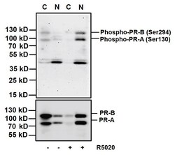
- Western blot analysis of Phosphorylated Progesterone Receptor (upper panel) and total Progesterone Receptor (lower panel) was performed by loading 20 µg of nuclear (N) or cytoplasmic (C) T47D cell lysates, untreated (-) or stimulated (+) with 100 nm promegestone (R5020) for 1 hour and 10 µL PageRuler Plus Prestained Protein Ladder (Product # 26619) per well onto a 4-20% Tris-Glycine polyacrylamide gel. Proteins were transferred to a nitrocellulose membrane using the G2 Fast Blotter (Product # 62288) and blocked with 5% Milk/TBST for at least 1 hour at room temperature. Phosphorylated Progesterone Receptor was detected using a Phospho-Progesterone Receptor (Ser294) mouse monoclonal antibody (upper panel, Product # MA1-414) at a concentration of 10 µg/mL and Total Progesterone Receptor was detected using a Progesterone Receptor mouse monoclonal antibody (lower panel, Product # MA1-12626) at a concentration of 1 µg/mL in blocking buffer overnight at 4°C on a rocking platform, followed by a Superclonal goat anti-Mouse IgG-HRP secondary antibody (Product # A28177) at a dilution of 1:2,000 for at least 1 hour at room temperature. Chemiluminescent detection was performed using SuperSignal West Pico (Product # 34078) and the myECL Imager (Product # 62236).
Supportive validation
- Submitted by
- Invitrogen Antibodies (provider)
- Main image
- Experimental details
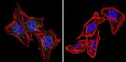
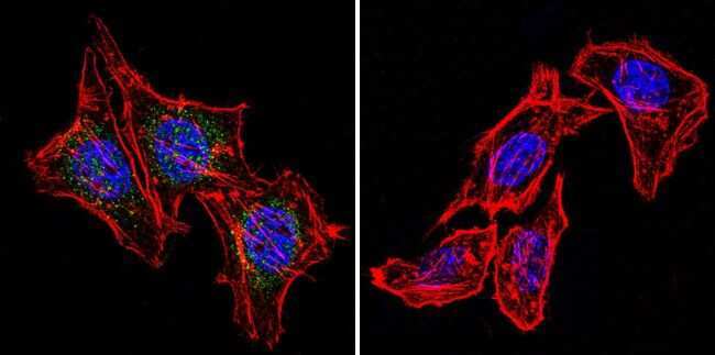
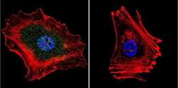
- Immunofluorescent analysis of Phosphorylated Progesterone Receptor using Phospho-Progesterone Receptor pSer294 Monoclonal Antibody (608) (Product # MA1-414) shows staining in Hela Cells. Phospho-Progesterone Receptor pSer294 (green), F-Actin staining with Phalloidin (red) and nuclei with DAPI (blue) is shown. Cells were grown on chamber slides and fixed with formaldehyde prior to staining. Cells were probed without (control) or with an antibody recognizing Phospho-Progesterone Receptor pSer294 (Product # MA1-414) at a dilution of 1:20 over night at 4 °C, washed with PBS and incubated with a DyLight-488 conjugated secondary antibody (Product # 35552 for GAR, Product # 35503 for GAM). Images were taken at 60X magnification.
- Submitted by
- Invitrogen Antibodies (provider)
- Main image

- Experimental details
- Immunofluorescent analysis of Phosphorylated Progesterone Receptor using Phospho-Progesterone Receptor pSer294 Monoclonal Antibody (608) (Product # MA1-414) shows staining in U251 Cells. Phospho-Progesterone Receptor pSer294 (green), F-Actin staining with Phalloidin (red) and nuclei with DAPI (blue) is shown. Cells were grown on chamber slides and fixed with formaldehyde prior to staining. Cells were probed without (control) or with an antibody recognizing Phospho-Progesterone Receptor pSer294 (Product # MA1-414) at a dilution of 1:20 over night at 4 °C, washed with PBS and incubated with a DyLight-488 conjugated secondary antibody (Product # 35552 for GAR, Product # 35503 for GAM). Images were taken at 60X magnification.
- Submitted by
- Invitrogen Antibodies (provider)
- Main image
- Experimental details
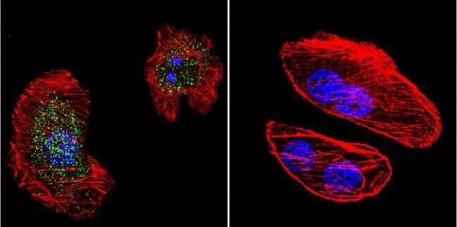
- Immunofluorescent analysis of Phosphorylated Progesterone Receptor using Phospho-Progesterone Receptor pSer294 Monoclonal Antibody (608) (Product # MA1-414) shows staining in MCF-7 Cells. Phospho-Progesterone Receptor pSer294 (green), F-Actin staining with Phalloidin (red) and nuclei with DAPI (blue) is shown. Cells were grown on chamber slides and fixed with formaldehyde prior to staining. Cells were probed without (control) or with an antibody recognizing Phospho-Progesterone Receptor pSer294 (Product # MA1-414) at a dilution of 1:20 over night at 4 °C, washed with PBS and incubated with a DyLight-488 conjugated secondary antibody (Product # 35552 for GAR, Product # 35503 for GAM). Images were taken at 60X magnification.

Supportive validation
- Submitted by
- Invitrogen Antibodies (provider)
- Main image
- Experimental details
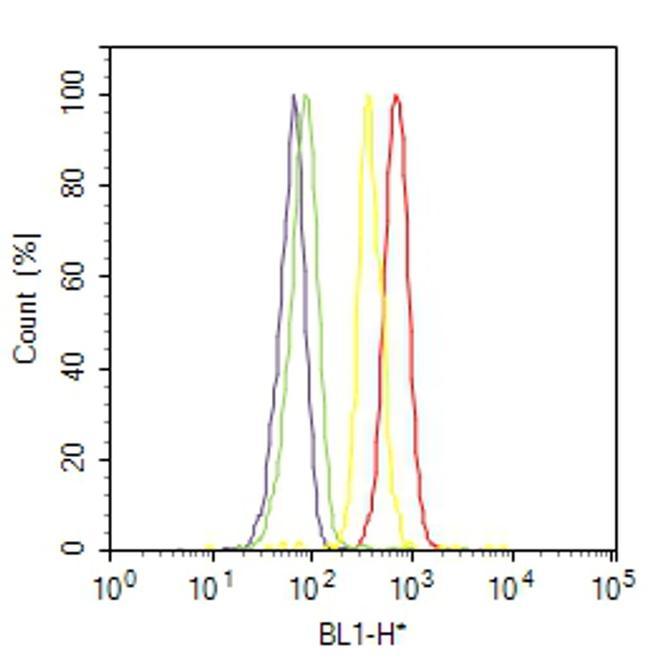
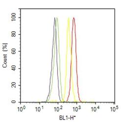
- Flow cytometry analysis of Progesterone [pSer294] was done on MCF7 cells. Cells were fixed with 70% ethanol for 10 minutes, permeabilized with 0.25% Triton™ X-100 for 20 minutes, and blocked with 5% BSA for 30 minutes at room temperature. Cells were labeled with Progesterone [pSer294] Mouse Monoclonal Antibody (MA1414, red histogram) or with mouse isotype control (yellow histogram) at 3-5 ug/million cells in 2.5% BSA. After incubation at room temperature for 2 hours, the cells were labeled with Alexa Fluor® 488 Rabbit Anti-Mouse Secondary Antibody (A11059) at a dilution of 1:400 for 30 minutes at room temperature. The representative 10,000 cells were acquired and analyzed for each sample using an Attune® Acoustic Focusing Cytometer. The purple histogram represents unstained control cells and the green histogram represents no-primary-antibody control.