 Explore
 Validate
 Learn
Learn Western blot
Antibody data
- Antibody Data
- Antigen structure
- References [4]
- Comments [0]
- Validations
- Western blot [3]
- Immunocytochemistry [2]
- Immunohistochemistry [1]
Submit
Validation data
Reference
Comment
Report error
- Product number
- MA5-14842 - Provider product page

- Provider
- Invitrogen Antibodies
- Product name
- Progesterone Receptor Monoclonal Antibody (R.809.9)
- Antibody type
- Monoclonal
- Antigen
- Synthetic peptide
- Description
- It is not recommended to aliquot this antibody.
- Antibody clone number
- R.809.9
- Concentration
- 243 µg/mL
Submitted references Progesterone Receptor Isoforms, Nuclear Corepressor-1 and Steroid Receptor Coactivator-1 and B-Cell Lymphoma 2 and Akt and Akt Phosphorylation Status in Uterine Myomas after Ulipristal Acetate Treatment: A Systematic Immunohistochemical Evaluation.
Tissue expression of steroid hormone receptors is associated with differential immune responsiveness.
TNF superfamily gene polymorphism as prognostic factor in early breast cancer.
Epidermal growth factor receptor signaling enhanced by long-term medroxyprogesterone acetate treatment in endometrial carcinoma.
Courtoy GE, Donnez J, Marbaix E, Barreira M, Luyckx M, Dolmans MM
Gynecologic and obstetric investigation 2018;83(5):443-454
Gynecologic and obstetric investigation 2018;83(5):443-454
Tissue expression of steroid hormone receptors is associated with differential immune responsiveness.
Butts CL, Jones YL, Lim JK, Salter CE, Belyavskaya E, Sternberg EM
Brain, behavior, and immunity 2011 Jul;25(5):1000-7
Brain, behavior, and immunity 2011 Jul;25(5):1000-7
TNF superfamily gene polymorphism as prognostic factor in early breast cancer.
Jung JH, Chae YS, Moon JH, Kang BW, Kim JG, Sohn SK, Park JY, Lee MH, Park HY
Journal of cancer research and clinical oncology 2010 May;136(5):685-94
Journal of cancer research and clinical oncology 2010 May;136(5):685-94
Epidermal growth factor receptor signaling enhanced by long-term medroxyprogesterone acetate treatment in endometrial carcinoma.
Zhao S, Chen X, Lu X, Yu Y, Feng Y
Gynecologic oncology 2007 Apr;105(1):45-54
Gynecologic oncology 2007 Apr;105(1):45-54
No comments: Submit comment
Supportive validation
- Submitted by
- Invitrogen Antibodies (provider)
- Main image
- Experimental details
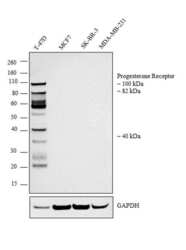
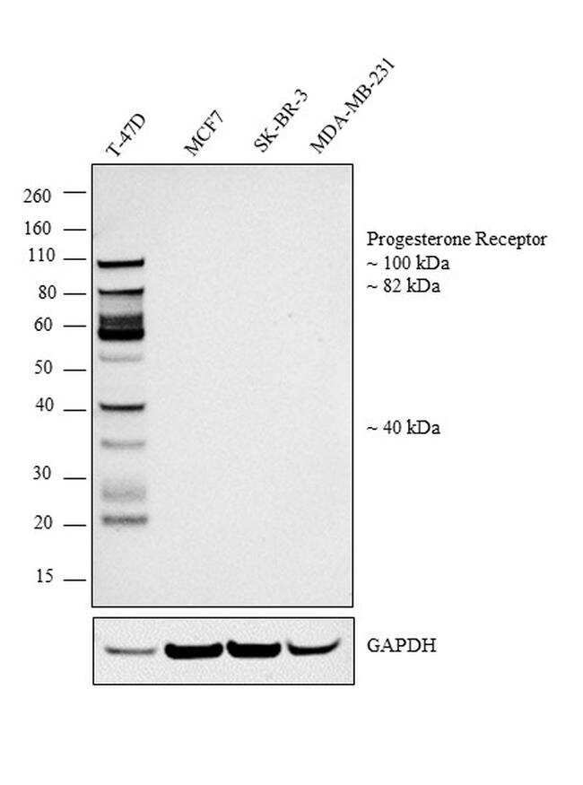
- Western blot analysis was performed on modified whole cell extracts (1% SDS) (30 µg lysate) of T-47D (Lane 1), MCF7 (Lane 2), SK-BR-3 (Lane3) and MDA-MB-231 (Lane 4). The blot was probed with Anti-Progesterone Receptor Monoclonal Antibody (R.809.9) (Product # MA5-14842, 1:1000 dilution) and detected by chemiluminescence using Goat anti-Rabbit IgG (H+L) Superclonal™ Secondary Antibody, HRP conjugate (Product # A27036, 0.25 µg/mL, 1:4000 dilution). Bands corresponding to multiple isoforms of Progesterone Receptor at 100, 82, and 40 kDa were detected in T-47D and undetected in low expressing MCF7, SK-BR-3 and MDA-MB-231 cell lines.
- Submitted by
- Invitrogen Antibodies (provider)
- Main image
- Experimental details
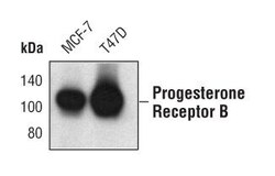
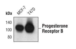
- Western blot analysis of Progesterone Receptor B in extracts from MCF-7 and T47D cells using Progesterone Receptor B monoclonal antibody (Product # MA5-14842).
- Submitted by
- Invitrogen Antibodies (provider)
- Main image
- Experimental details
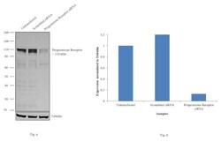
- Knockdown of Progesterone Receptor was achieved by transfecting T-47D with Progesterone Receptor specific siRNAs (Silencer® select Product # s10416; s10415). Western blot analysis (Fig. a) was performed using whole cell extracts from the Progesterone Receptor knockdown cells (lane 3), non-specific scrambled siRNA transfected cells (lane 2) and untransfected cells (lane 1). The blots were probed with Progesterone Receptor Polyclonal Antibody (Product # MA5-14842, 1:1000 dilution) and Goat anti-Mouse IgG (H+L) Superclonal™ Secondary Antibody, HRP conjugate (Product # A28177, 0.25 µg/mL 1:4000 dilution). Densitometric analysis of this western blot is shown in histogram (Fig. b). Decrease in signal upon siRNA mediated knock down confirms that antibody is specific to Progesterone Receptor.

Supportive validation
- Submitted by
- Invitrogen Antibodies (provider)
- Main image
- Experimental details
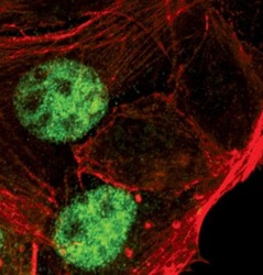
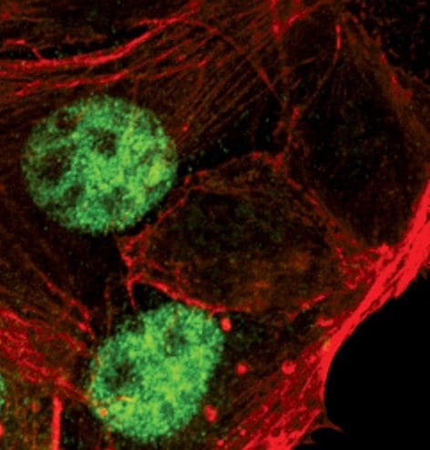
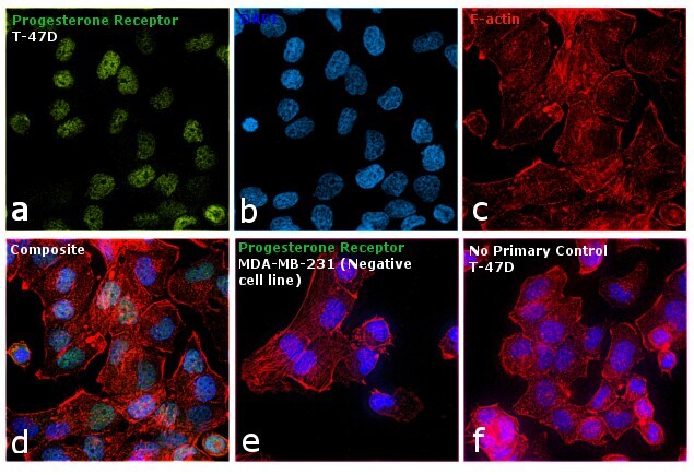
- Immunofluorescent analysis of Progesterone Receptor using a monoclonal antibody (Product # MA5-14842).
- Submitted by
- Invitrogen Antibodies (provider)
- Main image
- Experimental details
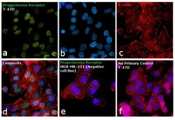
- Immunofluorescence analysis of Progesterone Receptor was performed using 70% confluent log phase T-47D cells. The cells were fixed with 4% paraformaldehyde for 10 minutes, permeabilized with 0.1% Triton™ X-100 for 15 minutes, and blocked with 1% BSA for 1 hour at room temperature. The cells were labeled with Progesterone Receptor Rabbit Monoclonal Antibody (R.809.9) (Product # MA5-14842) at 1:200 dilution in 0.1% BSA, incubated at 4 degree Celsius overnight and then labeled with Goat anti-Rabbit IgG (H+L) Superclonal™ Secondary Antibody, Alexa Fluor® 488 conjugate (Product # A27034) at a dilution of 1:2000 for 45 minutes at room temperature (Panel a: green). Nuclei (Panel b: blue) were stained with ProLong™ Diamond Antifade Mountant with DAPI (Product # P36962). F-actin (Panel c: red) was stained with Rhodamine Phalloidin (Product # R415). Panel d represents the merged image showing Nuclear localization. Panel e represents the negative cell line, MDA-MB-231 cells. Panel f represents control cells with no primary antibody to assess background. The images were captured at 60X magnification.
Supportive validation
- Submitted by
- Invitrogen Antibodies (provider)
- Main image
- Experimental details
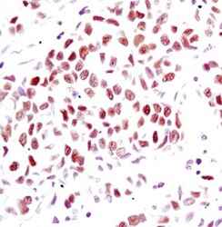
- Immunohistochemical analysis of Progesterone Receptor B in paraffin-embedded human breast carcinoma using a Progesterone Receptor B monoclonal antibody (Product # MA5-14842) in the presence of control peptide (left) or antigen-specific peptide (right).
