Explore
 Validate
 Learn
Learn Western blot
Antibody data
- Antibody Data
- Antigen structure
- References [39]
- Comments [0]
- Validations
- Western blot [2]
- Immunocytochemistry [2]
- Other assay [2]
Submit
Validation data
Reference
Comment
Report error
- Product number
- MA5-12658 - Provider product page

- Provider
- Invitrogen Antibodies
- Product name
- Progesterone Receptor Monoclonal Antibody (hPRa 7)
- Antibody type
- Monoclonal
- Antigen
- Other
- Description
- MA5-12658 targets Progesterone Receptor in IF, IP, and WB applications and shows reactivity with Human, mouse, Non-human primate, and Rat samples.
- Antibody clone number
- hPRa 7
- Concentration
- 0.2 mg/mL
Submitted references Mifepristone Treatment Promotes Testicular Leydig Cell Tumor Progression in Transgenic Mice.
Molecular mechanisms underlying mifepristone's agonistic action on ovarian cancer progression.
Tissue-specific progesterone receptor-chromatin binding and the regulation of progesterone-dependent gene expression.
Progesterone Receptors in Prostate Cancer: Progesterone receptor B is the isoform associated with disease progression.
RANK Signaling Amplifies WNT-Responsive Mammary Progenitors through R-SPONDIN1.
Tip30 controls differentiation of murine mammary luminal progenitor to estrogen receptor-positive luminal cell through regulating FoxA1 expression.
Amphiregulin mediates progesterone-induced mammary ductal development during puberty.
Expansion of CD11b(+)Ly6G (+)Ly6C (int) cells driven by medroxyprogesterone acetate in mice bearing breast tumors restrains NK cell effector functions.
Antiprogestin mifepristone inhibits the growth of cancer cells of reproductive and non-reproductive origin regardless of progesterone receptor expression.
Novel role of signal transducer and activator of transcription 3 as a progesterone receptor coactivator in breast cancer.
Age-related changes in the epithelial and stromal compartments of the mammary gland in normocalcemic mice lacking the vitamin D3 receptor.
Classical membrane progesterone receptors in murine mammary carcinomas: agonistic effects of progestins and RU-486 mediating rapid non-genomic effects.
Hypermethylation of the progesterone receptor A in constitutive antiprogestin-resistant mouse mammary carcinomas.
Inhibition of mammary tumor growth by estrogens: is there a specific role for estrogen receptors alpha and beta?
Amphiregulin mediates estrogen, progesterone, and EGFR signaling in the normal rat mammary gland and in hormone-dependent rat mammary cancers.
Progesterone receptor induces ErbB-2 nuclear translocation to promote breast cancer growth via a novel transcriptional effect: ErbB-2 function as a coactivator of Stat3.
Tip30 deletion in MMTV-Neu mice leads to enhanced EGFR signaling and development of estrogen receptor-positive and progesterone receptor-negative mammary tumors.
Catechol-o-methyltransferase expression and 2-methoxyestradiol affect microtubule dynamics and modify steroid receptor signaling in leiomyoma cells.
Activation of Stat3 by heregulin/ErbB-2 through the co-option of progesterone receptor signaling drives breast cancer growth.
Prognosis of uterine corpus cancer after tamoxifen treatment for breast cancer.
Carcinoma-associated fibroblasts activate progesterone receptors and induce hormone independent mammary tumor growth: A role for the FGF-2/FGFR-2 axis.
NK cells expressing a progesterone receptor are susceptible to progesterone-induced apoptosis.
Gene expression programs of human smooth muscle cells: tissue-specific differentiation and prognostic significance in breast cancers.
Progesterone receptor isoforms and proliferation in the rat mammary gland during development.
Expression profiles of sex steroid receptors in desmoid tumors.
Expression profiles of sex steroid receptors in desmoid tumors.
Progestin-induced caveolin-1 expression mediates breast cancer cell proliferation.
Phosphorylation of estrogen receptor alpha serine 167 is predictive of response to endocrine therapy and increases postrelapse survival in metastatic breast cancer.
Progesterone receptor isoform (A/B) ratio of human fetal membranes increases during term parturition.
Disappearance of steroid hormone dependency during malignant transformation of ovarian clear cell cancer.
Progestins induce transcriptional activation of signal transducer and activator of transcription 3 (Stat3) via a Jak- and Src-dependent mechanism in breast cancer cells.
Cytologic scoring of endometrioid adenocarcinoma of the endometrium.
Reduction of progesterone receptor expression in human cumulus cells at the time of oocyte collection during IVF is associated with good embryo quality.
Inhibition of in vivo breast cancer growth by antisense oligodeoxynucleotides to type I insulin-like growth factor receptor mRNA involves inactivation of ErbBs, PI-3K/Akt and p42/p44 MAPK signaling pathways but not modulation of progesterone receptor activity.
Nuclear receptor DAX-1 in human common epithelial ovarian carcinoma: an independent prognostic factor of clinical outcome.
Nuclear receptor DAX-1 in human common epithelial ovarian carcinoma: an independent prognostic factor of clinical outcome.
Medroxyprogesterone acetate enhances in vivo and in vitro antibody production.
Overexpression of aromatase leads to hyperplasia and changes in the expression of genes involved in apoptosis, cell cycle, growth, and tumor suppressor functions in the mammary glands of transgenic mice.
Identification of progesterone receptor in human subcutaneous adipose tissue.
Ponikwicka-Tyszko D, Chrusciel M, Pulawska K, Bernaczyk P, Sztachelska M, Guo P, Li X, Toppari J, Huhtaniemi IT, Wołczyński S, Rahman NA
Cancers 2020 Nov 4;12(11)
Cancers 2020 Nov 4;12(11)
Molecular mechanisms underlying mifepristone's agonistic action on ovarian cancer progression.
Ponikwicka-Tyszko D, Chrusciel M, Stelmaszewska J, Bernaczyk P, Chrusciel P, Sztachelska M, Scheinin M, Bidzinski M, Szamatowicz J, Huhtaniemi IT, Wolczynski S, Rahman NA
EBioMedicine 2019 Sep;47:170-183
EBioMedicine 2019 Sep;47:170-183
Tissue-specific progesterone receptor-chromatin binding and the regulation of progesterone-dependent gene expression.
Dinh DT, Breen J, Akison LK, DeMayo FJ, Brown HM, Robker RL, Russell DL
Scientific reports 2019 Aug 19;9(1):11966
Scientific reports 2019 Aug 19;9(1):11966
Progesterone Receptors in Prostate Cancer: Progesterone receptor B is the isoform associated with disease progression.
Grindstad T, Richardsen E, Andersen S, Skjefstad K, Rakaee Khanehkenari M, Donnem T, Ness N, Nordby Y, Bremnes RM, Al-Saad S, Busund LT
Scientific reports 2018 Jul 27;8(1):11358
Scientific reports 2018 Jul 27;8(1):11358
RANK Signaling Amplifies WNT-Responsive Mammary Progenitors through R-SPONDIN1.
Joshi PA, Waterhouse PD, Kannan N, Narala S, Fang H, Di Grappa MA, Jackson HW, Penninger JM, Eaves C, Khokha R
Stem cell reports 2015 Jul 14;5(1):31-44
Stem cell reports 2015 Jul 14;5(1):31-44
Tip30 controls differentiation of murine mammary luminal progenitor to estrogen receptor-positive luminal cell through regulating FoxA1 expression.
Chen F, Li A, Gao S, Hollern D, Williams M, Liu F, VanSickle EA, Andrechek E, Zhang C, Yang C, Luo R, Xiao H
Cell death & disease 2014 May 22;5(5):e1242
Cell death & disease 2014 May 22;5(5):e1242
Amphiregulin mediates progesterone-induced mammary ductal development during puberty.
Aupperlee MD, Leipprandt JR, Bennett JM, Schwartz RC, Haslam SZ
Breast cancer research : BCR 2013 May 25;15(3):R44
Breast cancer research : BCR 2013 May 25;15(3):R44
Expansion of CD11b(+)Ly6G (+)Ly6C (int) cells driven by medroxyprogesterone acetate in mice bearing breast tumors restrains NK cell effector functions.
Spallanzani RG, Dalotto-Moreno T, Raffo Iraolagoitia XL, Ziblat A, Domaica CI, Avila DE, Rossi LE, Fuertes MB, Battistone MA, Rabinovich GA, Salatino M, Zwirner NW
Cancer immunology, immunotherapy : CII 2013 Dec;62(12):1781-95
Cancer immunology, immunotherapy : CII 2013 Dec;62(12):1781-95
Antiprogestin mifepristone inhibits the growth of cancer cells of reproductive and non-reproductive origin regardless of progesterone receptor expression.
Tieszen CR, Goyeneche AA, Brandhagen BN, Ortbahn CT, Telleria CM
BMC cancer 2011 May 27;11:207
BMC cancer 2011 May 27;11:207
Novel role of signal transducer and activator of transcription 3 as a progesterone receptor coactivator in breast cancer.
Proietti CJ, Béguelin W, Flaqué MC, Cayrol F, Rivas MA, Tkach M, Charreau EH, Schillaci R, Elizalde PV
Steroids 2011 Mar;76(4):381-92
Steroids 2011 Mar;76(4):381-92
Age-related changes in the epithelial and stromal compartments of the mammary gland in normocalcemic mice lacking the vitamin D3 receptor.
Welsh J, Zinser LN, Mianecki-Morton L, Martin J, Waltz SE, James H, Zinser GM
PloS one 2011 Jan 26;6(1):e16479
PloS one 2011 Jan 26;6(1):e16479
Classical membrane progesterone receptors in murine mammary carcinomas: agonistic effects of progestins and RU-486 mediating rapid non-genomic effects.
Bottino MC, Cerliani JP, Rojas P, Giulianelli S, Soldati R, Mondillo C, Gorostiaga MA, Pignataro OP, Calvo JC, Gutkind JS, Panomwat Amornphimoltham, Molinolo AA, Lüthy IA, Lanari C
Breast cancer research and treatment 2011 Apr;126(3):621-36
Breast cancer research and treatment 2011 Apr;126(3):621-36
Hypermethylation of the progesterone receptor A in constitutive antiprogestin-resistant mouse mammary carcinomas.
Wargon V, Fernandez SV, Goin M, Giulianelli S, Russo J, Lanari C
Breast cancer research and treatment 2011 Apr;126(2):319-32
Breast cancer research and treatment 2011 Apr;126(2):319-32
Inhibition of mammary tumor growth by estrogens: is there a specific role for estrogen receptors alpha and beta?
Soldati R, Wargon V, Cerliani JP, Giulianelli S, Vanzulli SI, Gorostiaga MA, Bolado J, do Campo P, Molinolo A, Vollmer G, Lanari C
Breast cancer research and treatment 2010 Oct;123(3):709-24
Breast cancer research and treatment 2010 Oct;123(3):709-24
Amphiregulin mediates estrogen, progesterone, and EGFR signaling in the normal rat mammary gland and in hormone-dependent rat mammary cancers.
Kariagina A, Xie J, Leipprandt JR, Haslam SZ
Hormones & cancer 2010 Oct;1(5):229-44
Hormones & cancer 2010 Oct;1(5):229-44
Progesterone receptor induces ErbB-2 nuclear translocation to promote breast cancer growth via a novel transcriptional effect: ErbB-2 function as a coactivator of Stat3.
Béguelin W, Díaz Flaqué MC, Proietti CJ, Cayrol F, Rivas MA, Tkach M, Rosemblit C, Tocci JM, Charreau EH, Schillaci R, Elizalde PV
Molecular and cellular biology 2010 Dec;30(23):5456-72
Molecular and cellular biology 2010 Dec;30(23):5456-72
Tip30 deletion in MMTV-Neu mice leads to enhanced EGFR signaling and development of estrogen receptor-positive and progesterone receptor-negative mammary tumors.
Zhang C, Mori M, Gao S, Li A, Hoshino I, Aupperlee MD, Haslam SZ, Xiao H
Cancer research 2010 Dec 15;70(24):10224-33
Cancer research 2010 Dec 15;70(24):10224-33
Catechol-o-methyltransferase expression and 2-methoxyestradiol affect microtubule dynamics and modify steroid receptor signaling in leiomyoma cells.
Salama SA, Kamel MW, Botting S, Salih SM, Borahay MA, Hamed AA, Kilic GS, Saeed M, Williams MY, Diaz-Arrastia CR
PloS one 2009 Oct 7;4(10):e7356
PloS one 2009 Oct 7;4(10):e7356
Activation of Stat3 by heregulin/ErbB-2 through the co-option of progesterone receptor signaling drives breast cancer growth.
Proietti CJ, Rosemblit C, Beguelin W, Rivas MA, Díaz Flaqué MC, Charreau EH, Schillaci R, Elizalde PV
Molecular and cellular biology 2009 Mar;29(5):1249-65
Molecular and cellular biology 2009 Mar;29(5):1249-65
Prognosis of uterine corpus cancer after tamoxifen treatment for breast cancer.
Hoogendoorn WE, Hollema H, van Boven HH, Bergman E, de Leeuw-Mantel G, Platteel I, Fles R, Nederlof PM, Mourits MJ, van Leeuwen FE, Comprehensive Cancer Centers TAMARISK-group
Breast cancer research and treatment 2008 Nov;112(1):99-108
Breast cancer research and treatment 2008 Nov;112(1):99-108
Carcinoma-associated fibroblasts activate progesterone receptors and induce hormone independent mammary tumor growth: A role for the FGF-2/FGFR-2 axis.
Giulianelli S, Cerliani JP, Lamb CA, Fabris VT, Bottino MC, Gorostiaga MA, Novaro V, Góngora A, Baldi A, Molinolo A, Lanari C
International journal of cancer 2008 Dec 1;123(11):2518-31
International journal of cancer 2008 Dec 1;123(11):2518-31
NK cells expressing a progesterone receptor are susceptible to progesterone-induced apoptosis.
Arruvito L, Giulianelli S, Flores AC, Paladino N, Barboza M, Lanari C, Fainboim L
Journal of immunology (Baltimore, Md. : 1950) 2008 Apr 15;180(8):5746-53
Journal of immunology (Baltimore, Md. : 1950) 2008 Apr 15;180(8):5746-53
Gene expression programs of human smooth muscle cells: tissue-specific differentiation and prognostic significance in breast cancers.
Chi JT, Rodriguez EH, Wang Z, Nuyten DS, Mukherjee S, van de Rijn M, van de Vijver MJ, Hastie T, Brown PO
PLoS genetics 2007 Sep;3(9):1770-84
PLoS genetics 2007 Sep;3(9):1770-84
Progesterone receptor isoforms and proliferation in the rat mammary gland during development.
Kariagina A, Aupperlee MD, Haslam SZ
Endocrinology 2007 Jun;148(6):2723-36
Endocrinology 2007 Jun;148(6):2723-36
Expression profiles of sex steroid receptors in desmoid tumors.
Ishizuka M, Hatori M, Dohi O, Suzuki T, Miki Y, Tazawa C, Sasano H, Kokubun S
The Tohoku journal of experimental medicine 2006 Nov;210(3):189-98
The Tohoku journal of experimental medicine 2006 Nov;210(3):189-98
Expression profiles of sex steroid receptors in desmoid tumors.
Ishizuka M, Hatori M, Dohi O, Suzuki T, Miki Y, Tazawa C, Sasano H, Kokubun S
The Tohoku journal of experimental medicine 2006 Nov;210(3):189-98
The Tohoku journal of experimental medicine 2006 Nov;210(3):189-98
Progestin-induced caveolin-1 expression mediates breast cancer cell proliferation.
Salatino M, Beguelin W, Peters MG, Carnevale R, Proietti CJ, Galigniana MD, Vedoy CG, Schillaci R, Charreau EH, Sogayar MC, Elizalde PV
Oncogene 2006 Dec 14;25(59):7723-39
Oncogene 2006 Dec 14;25(59):7723-39
Phosphorylation of estrogen receptor alpha serine 167 is predictive of response to endocrine therapy and increases postrelapse survival in metastatic breast cancer.
Yamashita H, Nishio M, Kobayashi S, Ando Y, Sugiura H, Zhang Z, Hamaguchi M, Mita K, Fujii Y, Iwase H
Breast cancer research : BCR 2005;7(5):R753-64
Breast cancer research : BCR 2005;7(5):R753-64
Progesterone receptor isoform (A/B) ratio of human fetal membranes increases during term parturition.
Oh SY, Kim CJ, Park I, Romero R, Sohn YK, Moon KC, Yoon BH
American journal of obstetrics and gynecology 2005 Sep;193(3 Pt 2):1156-60
American journal of obstetrics and gynecology 2005 Sep;193(3 Pt 2):1156-60
Disappearance of steroid hormone dependency during malignant transformation of ovarian clear cell cancer.
Akahane T, Sekizawa A, Okuda T, Kushima M, Saito H, Okai T
International journal of gynecological pathology : official journal of the International Society of Gynecological Pathologists 2005 Oct;24(4):369-76
International journal of gynecological pathology : official journal of the International Society of Gynecological Pathologists 2005 Oct;24(4):369-76
Progestins induce transcriptional activation of signal transducer and activator of transcription 3 (Stat3) via a Jak- and Src-dependent mechanism in breast cancer cells.
Proietti C, Salatino M, Rosemblit C, Carnevale R, Pecci A, Kornblihtt AR, Molinolo AA, Frahm I, Charreau EH, Schillaci R, Elizalde PV
Molecular and cellular biology 2005 Jun;25(12):4826-40
Molecular and cellular biology 2005 Jun;25(12):4826-40
Cytologic scoring of endometrioid adenocarcinoma of the endometrium.
Nishimura Y, Watanabe J, Jobo T, Hattori M, Arai T, Kuramoto H
Cancer 2005 Feb 25;105(1):8-12
Cancer 2005 Feb 25;105(1):8-12
Reduction of progesterone receptor expression in human cumulus cells at the time of oocyte collection during IVF is associated with good embryo quality.
Hasegawa J, Yanaihara A, Iwasaki S, Otsuka Y, Negishi M, Akahane T, Okai T
Human reproduction (Oxford, England) 2005 Aug;20(8):2194-200
Human reproduction (Oxford, England) 2005 Aug;20(8):2194-200
Inhibition of in vivo breast cancer growth by antisense oligodeoxynucleotides to type I insulin-like growth factor receptor mRNA involves inactivation of ErbBs, PI-3K/Akt and p42/p44 MAPK signaling pathways but not modulation of progesterone receptor activity.
Salatino M, Schillaci R, Proietti CJ, Carnevale R, Frahm I, Molinolo AA, Iribarren A, Charreau EH, Elizalde PV
Oncogene 2004 Jul 1;23(30):5161-74
Oncogene 2004 Jul 1;23(30):5161-74
Nuclear receptor DAX-1 in human common epithelial ovarian carcinoma: an independent prognostic factor of clinical outcome.
Abd-Elaziz M, Akahira J, Moriya T, Suzuki T, Yaegashi N, Sasano H
Cancer science 2003 Nov;94(11):980-5
Cancer science 2003 Nov;94(11):980-5
Nuclear receptor DAX-1 in human common epithelial ovarian carcinoma: an independent prognostic factor of clinical outcome.
Abd-Elaziz M, Akahira J, Moriya T, Suzuki T, Yaegashi N, Sasano H
Cancer science 2003 Nov;94(11):980-5
Cancer science 2003 Nov;94(11):980-5
Medroxyprogesterone acetate enhances in vivo and in vitro antibody production.
Vermeulen M, Pazos P, Lanari C, Molinolo A, Gamberale R, Geffner JR, Giordano M
Immunology 2001 Sep;104(1):80-6
Immunology 2001 Sep;104(1):80-6
Overexpression of aromatase leads to hyperplasia and changes in the expression of genes involved in apoptosis, cell cycle, growth, and tumor suppressor functions in the mammary glands of transgenic mice.
Kirma N, Gill K, Mandava U, Tekmal RR
Cancer research 2001 Mar 1;61(5):1910-8
Cancer research 2001 Mar 1;61(5):1910-8
Identification of progesterone receptor in human subcutaneous adipose tissue.
O'Brien SN, Welter BH, Mantzke KA, Price TM
The Journal of clinical endocrinology and metabolism 1998 Feb;83(2):509-13
The Journal of clinical endocrinology and metabolism 1998 Feb;83(2):509-13
No comments: Submit comment
Supportive validation
- Submitted by
- Invitrogen Antibodies (provider)
- Main image

- Experimental details
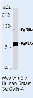
- Western blot of Progesterone Receptor using Progesterone Receptor Monoclonal Antibody (Product # MA5-12658) on Human Breast Cancer Tissue.
- Submitted by
- Invitrogen Antibodies (provider)
- Main image

- Experimental details
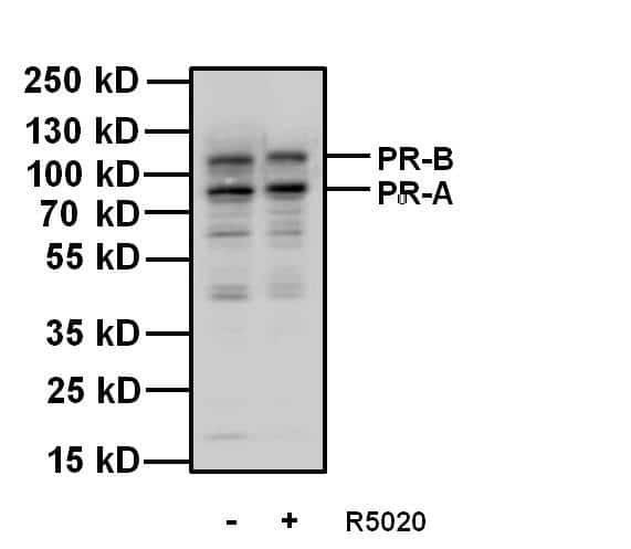
- Western blot analysis of Progesterone Receptor was performed by loading 20 µg of T47D cell lysates, untreated (-) or stimulated (+) with 100 nm promegestone (R5020) for 1 hour and 10 µL PageRuler Plus Prestained Protein Ladder (Product # 26619) per well onto a 4-20% Tris-Glycine polyacrylamide gel. Proteins were transferred to a nitrocellulose membrane using the G2 Fast Blotter (Product # 62288) and blocked with 5% Milk/TBST for at least 1 hour at room temperature. Progesterone Receptor was detected using a Progesterone Receptor mouse monoclonal antibody, Product # MA5-12658, at a concentration of 1 µg/mL in blocking buffer overnight at 4°C on a rocking platform, followed by a Superclonal goat anti-Mouse IgG-HRP secondary antibody (Product # A28177) at a dilution of 1:2,000 for at least 1 hour at room temperature. Chemiluminescent detection was performed using SuperSignal West Pico (Product # 34078) and the myECL Imager (Product # 62236).
Supportive validation
- Submitted by
- Invitrogen Antibodies (provider)
- Main image

- Experimental details
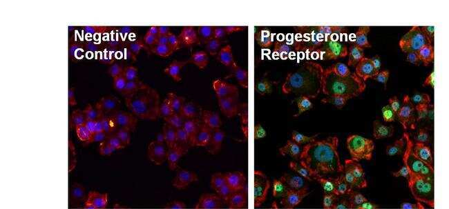
- Immunofluorescent analysis of Progesterone Receptor (green) in T47D cells. The cells were fixed with formalin for 15 minutes, permeabilized with 0.1% Triton X-100 in TBS for 10 minutes, and blocked with 3% Blocker BSA (Product # 37525) for 15 minutes at room temperature. Cells were stained with or without Progesterone Receptor mouse monoclonal antibody (Product # MA5-12658), at a concentration of 10 µg/mL for 1 hour at 37C, and then incubated with a Alexa Fluor 488 Superclonal goat anti-mouse IgG secondary antibody (Product # A28175) at a dilution of 1:1000 for 30 minutes at room temperature (both panels, green). Nuclei (both panels, blue) were stained with Hoechst 33342 dye (Product # 62249). Images were taken on a Thermo Scientific ToxInsight at 20X magnification.
- Submitted by
- Invitrogen Antibodies (provider)
- Main image

- Experimental details
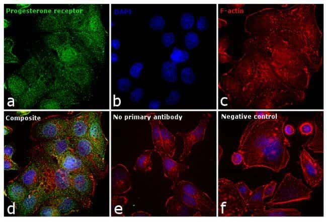
- Immunofluorescence analysis of Progesterone receptor was performed using 70% confluent log phase T-47D cells. The cells were fixed with 4% paraformaldehyde for 10 minutes, permeabilized with 0.1% Triton™ X-100 for 15 minutes, and blocked with 1% BSA for 1 hour at room temperature. The cells were labeled with Progesterone Receptor Monoclonal Antibody (hPRa 7) (Product # MA5-12658) at 1:50 dilution in 0.1% BSA, incubated at 4 degree celsius overnight and then labeled with Goat anti-Mouse IgG (H+L) Superclonal™ Secondary Antibody, Alexa Fluor® 488 conjugate (Product # A28175) at a dilution of 1:2000 for 45 minutes at room temperature (Panel a: green).Nuclei (Panel b: blue) were stained with SlowFade® Gold Antifade Mountant with DAPI (Product # S36938). F-actin (Panel c: red) was stained with Rhodamine Phalloidin (Product # R415, 1:300). Panel d represents the merged image showing cytoplasmic and nuclear localization. Panel e represents control cells with no primary antibody to assess background. Panel f shows the negative model, SK-BR-3, that does not express Progesterone receptor. The images were captured at 60X magnification.
Supportive validation
- Submitted by
- Invitrogen Antibodies (provider)
- Main image

- Experimental details
- Figure 3 Pgr gene profiling in MF- and P4-treated transgenic Inhalpha/Tag TG mice and BLTK-1 cells proliferation and invasion. qPCR analysis of Pgr ( A ), Pgrmc1 ( B ), Pgrmc2 ( C ), Paqr7 (mPRalpha) ( D ), Paqr8 (mPRbeta) ( E ), and Paqr5 (mPRgamma) ( F ) expression in the non-, MF- and P4-treated tumors of Inhalpha/Tag TG mice. Each bar represents the mean +- SEM relative to Ppia. Immunohistochemical staining of PGR in the control ( G ), MF-treated ( H ) and P4-treated ( I ) tumors and of PGRMC1 in control ( J ), MF-treated ( K ) and P4-treated ( L ) LCTs of Inhalpha/Tag TG mice. Scale bar, 100 mum. Asterisks indicate significant differences between the control and treated groups (**, p < 0.01). C, control; Inhalpha/Tag TG mice; transgenic mice expressing the SV40 Taq oncogene under the inhibin alpha promoter; LCT, Leydig cell tumor; MF, mifepristone; P4, progesterone.

- Submitted by
- Invitrogen Antibodies (provider)
- Main image

- Experimental details
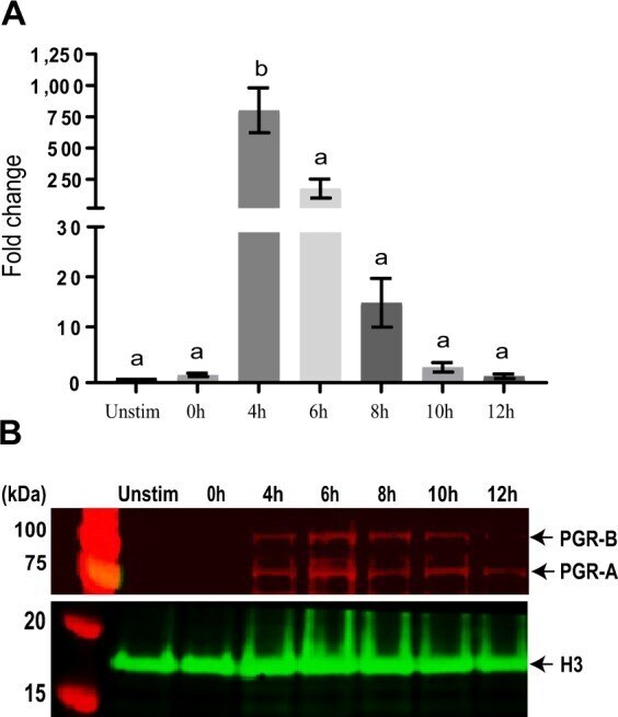
- Figure 1 PGR mRNA and protein are induced by the LH surge in granulosa cells. ( A ) PGR mRNA expression in eCG and hCG-primed granulosa cells at no eCG + hCG (unstim) and eCG + hCG 0-12 h post-hCG stimulation. RT-qPCR was performed on samples of 1-3 ovaries. N = 3 independent experiments, bars with different superscripts are significantly different, F 6,14 = 17.53, p < 0.0001 (one-way ANOVA). ( B ) Western blot of PGR in granulosa cells during ovulation (unstim or eCG + hCG 0-12 h post-hCG). Western blot was performed in biological triplicates, with 3-4 mice per time point in each replicate. One representative example of three highly similar results is shown. Full-length blot is available in Suppl. Data 3 .