 Explore
 Validate
 Learn
LearnMA1-071
antibody from Invitrogen Antibodies
Targeting: NUP62
DKFZp547L134, FLJ20822, FLJ43869, IBSN, MGC841, p62, SNDI
 Western blot
 Immunocytochemistry
Antibody data
- Antibody Data
- Antigen structure
- References [10]
- Comments [0]
- Validations
- Western blot [2]
- Immunohistochemistry [3]
Submit
Validation data
Reference
Comment
Report error
- Product number
- MA1-071 - Provider product page

- Provider
- Invitrogen Antibodies
- Product name
- Anti-NUP62 Monoclonal Antibody (RL1)
- Antibody type
- Monoclonal
- Antigen
- Purifed from natural sources
- Description
- MA1-071 detects nuclear pore-O-linked glycoprotein from rat, amphibian and yeast. MA1-071 has been successfully used in Western blot, immunofluorescence and immunoprecipitation procedures. By Western blot, this antibody detects up to eight different proteins from the nuclear pore complex (NPC) of approximately 210, 180, 145, 100, 63, 58, 54 and 45 kDa. Immunofluorescence staining of NPC O-linked glycoproteins with MA1-071 results in exclusive labeling of the NPC proteins on a wide variety of mammalian cells as well as S. cerevisiae and Xenopus. Labeling occurs exclusively at the NPC with most of the labeling at the cytoplasmic and nucleoplasmic margins. Microinjected MA1-071 inhibits both protein import and RNA export in Xenopus oocytes. The MA1-071 immunogen is pore complex-lamina fraction purified from rat liver nuclear envelopes.
- Reactivity
- Rat, Yeast
- Host
- Mouse
- Isotype
- IgM
- Antibody clone number
- RL1
- Vial size
- 100 µL
- Concentration
- Lot Dependent
- Storage
- -20° C, Avoid Freeze/Thaw Cycles
Submitted references Cellular and Molecular Features of Developmentally Programmed Genome Rearrangement in a Vertebrate (Sea Lamprey: Petromyzon marinus).
Dynamic plasticity of large-scale chromatin structure revealed by self-assembly of engineered chromosome regions.
Caenorhabditis elegans ortholog of a diabetes susceptibility locus: oga-1 (O-GlcNAcase) knockout impacts O-GlcNAc cycling, metabolism, and dauer.
Exportin-5, a novel karyopherin, mediates nuclear export of double-stranded RNA binding proteins.
Identification and characterization of Caenorhabditis elegans gamma-tubulin in dividing cells and differentiated tissues.
A role for RanBP1 in the release of CRM1 from the nuclear pore complex in a terminal step of nuclear export.
Lamin-binding fragment of LAP2 inhibits increase in nuclear volume during the cell cycle and progression into S phase.
A conditional allele of the novel repeat-containing yeast nucleoporin RAT7/NUP159 causes both rapid cessation of mRNA export and reversible clustering of nuclear pore complexes.
O-linked glycoproteins of the nuclear pore complex interact with a cytosolic factor required for nuclear protein import.
Nuclear protein import is inhibited by an antibody to a lumenal epitope of a nuclear pore complex glycoprotein.
Timoshevskiy VA, Herdy JR, Keinath MC, Smith JJ
PLoS genetics 2016 Jun;12(6):e1006103
PLoS genetics 2016 Jun;12(6):e1006103
Dynamic plasticity of large-scale chromatin structure revealed by self-assembly of engineered chromosome regions.
Sinclair P, Bian Q, Plutz M, Heard E, Belmont AS
The Journal of cell biology 2010 Sep 6;190(5):761-76
The Journal of cell biology 2010 Sep 6;190(5):761-76
Caenorhabditis elegans ortholog of a diabetes susceptibility locus: oga-1 (O-GlcNAcase) knockout impacts O-GlcNAc cycling, metabolism, and dauer.
Forsythe ME, Love DC, Lazarus BD, Kim EJ, Prinz WA, Ashwell G, Krause MW, Hanover JA
Proceedings of the National Academy of Sciences of the United States of America 2006 Aug 8;103(32):11952-7
Proceedings of the National Academy of Sciences of the United States of America 2006 Aug 8;103(32):11952-7
Exportin-5, a novel karyopherin, mediates nuclear export of double-stranded RNA binding proteins.
Brownawell AM, Macara IG
The Journal of cell biology 2002 Jan 7;156(1):53-64
The Journal of cell biology 2002 Jan 7;156(1):53-64
Identification and characterization of Caenorhabditis elegans gamma-tubulin in dividing cells and differentiated tissues.
Bobinnec Y, Fukuda M, Nishida E
Journal of cell science 2000 Nov;113 Pt 21:3747-59
Journal of cell science 2000 Nov;113 Pt 21:3747-59
A role for RanBP1 in the release of CRM1 from the nuclear pore complex in a terminal step of nuclear export.
Kehlenbach RH, Dickmanns A, Kehlenbach A, Guan T, Gerace L
The Journal of cell biology 1999 May 17;145(4):645-57
The Journal of cell biology 1999 May 17;145(4):645-57
Lamin-binding fragment of LAP2 inhibits increase in nuclear volume during the cell cycle and progression into S phase.
Yang L, Guan T, Gerace L
The Journal of cell biology 1997 Dec 1;139(5):1077-87
The Journal of cell biology 1997 Dec 1;139(5):1077-87
A conditional allele of the novel repeat-containing yeast nucleoporin RAT7/NUP159 causes both rapid cessation of mRNA export and reversible clustering of nuclear pore complexes.
Gorsch LC, Dockendorff TC, Cole CN
The Journal of cell biology 1995 May;129(4):939-55
The Journal of cell biology 1995 May;129(4):939-55
O-linked glycoproteins of the nuclear pore complex interact with a cytosolic factor required for nuclear protein import.
Sterne-Marr R, Blevitt JM, Gerace L
The Journal of cell biology 1992 Jan;116(2):271-80
The Journal of cell biology 1992 Jan;116(2):271-80
Nuclear protein import is inhibited by an antibody to a lumenal epitope of a nuclear pore complex glycoprotein.
Greber UF, Gerace L
The Journal of cell biology 1992 Jan;116(1):15-30
The Journal of cell biology 1992 Jan;116(1):15-30
No comments: Submit comment
Supportive validation
- Submitted by
- Invitrogen Antibodies (provider)
- Main image
- Experimental details
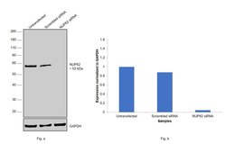
- Knockdown of NUP62 was achieved by transfecting HEK-293 with NUP62 specific siRNAs (Silencer® select Product # s24248). Western blot analysis (Fig. a) was performed using whole cell extracts from NUP62 knockdown cells (Lane 3), non-specific scrambled siRNA transfected cells (Lane 2) and untransfected cells (Lane 1). The blot was probed with NUP62 Monoclonal Antibody (RL1) (Product # MA1-071, 1:1000 dilution) and Goat anti-Mouse IgG (H+L), Superclonal™ Recombinant Secondary Antibody, HRP (Product # A28177, 1:4000 dilution). Densitometric analysis of this western blot is shown in histogram (Fig. b). Decrease in signal upon siRNA mediated knock down confirms that antibody is specific to NUP62.

- Submitted by
- Invitrogen Antibodies (provider)
- Main image
- Experimental details
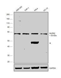
- Western blot was performed using NUP62 Monoclonal Antibody (RL1) (Product # MA1-071) and a 53kDa band corresponding to NUP62 was observed across cell lines along with an uncharacterized band (*) at ~45 kDa. Whole cell extracts (30 µg lysate) of HEK-293 (Lane 1), THP-1 (Lane 2), HeLa (Lane 3) and PC-12 (Lane 4) were electrophoresed using NuPAGE™ 10% Bis-Tris Protein Gel (Product # NP0302BOX). Resolved proteins were then transferred onto a nitrocellulose membrane (Product # IB23001) by iBlot® 2 Dry Blotting System (Product # IB21001). The blot was probed with the primary antibody (1:1000 dilution) and detected by chemiluminescence with Goat anti-Mouse IgG (H+L), Superclonal™ Recombinant Secondary Antibody, HRP (Product # A28177, 1:4000 dilution) using the iBright FL 1000 (Product # A32752). Chemiluminescent detection was performed using Novex® ECL Chemiluminescent Substrate Reagent Kit (Product # WP20005).

Supportive validation
- Submitted by
- Invitrogen Antibodies (provider)
- Main image

- Experimental details
- Immunohistochemistry was performed on normal biopsies of deparaffinized Rat brain tissue. To expose target proteins, heat induced antigen retrieval was performed using 10mM sodium citrate (pH6.0) buffer, microwaved for 8-15 minutes. Following antigen retrieval tissues were blocked in 3% BSA-PBS for 30 minutes at room temperature. Tissues were then probed at a dilution of 1:200 with a mouse monoclonal antibody recognizing Nuclear Pore-O-Linked Glycoprotein (Product # MA1-071) or without primary antibody (negative control) overnight at 4°C in a humidified chamber. Tissues were washed extensively with PBST and endogenous peroxidase activity was quenched with a peroxidase suppressor. Detection was performed using a biotin-conjugated secondary antibody and SA-HRP, followed by colorimetric detection using DAB. Tissues were counterstained with hematoxylin and prepped for mounting.

- Submitted by
- Invitrogen Antibodies (provider)
- Main image

- Experimental details
- Immunohistochemistry was performed on normal biopsies of deparaffinized Rat kidney tissue. To expose target proteins, heat induced antigen retrieval was performed using 10mM sodium citrate (pH6.0) buffer, microwaved for 8-15 minutes. Following antigen retrieval tissues were blocked in 3% BSA-PBS for 30 minutes at room temperature. Tissues were then probed at a dilution of 1:200 with a mouse monoclonal antibody recognizing Nuclear Pore-O-Linked Glycoprotein (Product # MA1-071) or without primary antibody (negative control) overnight at 4°C in a humidified chamber. Tissues were washed extensively with PBST and endogenous peroxidase activity was quenched with a peroxidase suppressor. Detection was performed using a biotin-conjugated secondary antibody and SA-HRP, followed by colorimetric detection using DAB. Tissues were counterstained with hematoxylin and prepped for mounting.

- Submitted by
- Invitrogen Antibodies (provider)
- Main image

- Experimental details
- Immunohistochemistry was performed on normal biopsies of deparaffinized Rat lymph node tissue. To expose target proteins, heat induced antigen retrieval was performed using 10mM sodium citrate (pH6.0) buffer, microwaved for 8-15 minutes. Following antigen retrieval tissues were blocked in 3% BSA-PBS for 30 minutes at room temperature. Tissues were then probed at a dilution of 1:200 with a mouse monoclonal antibody recognizing Nuclear Pore-O-Linked Glycoprotein (Product # MA1-071) or without primary antibody (negative control) overnight at 4°C in a humidified chamber. Tissues were washed extensively with PBST and endogenous peroxidase activity was quenched with a peroxidase suppressor. Detection was performed using a biotin-conjugated secondary antibody and SA-HRP, followed by colorimetric detection using DAB. Tissues were counterstained with hematoxylin and prepped for mounting.
