Explore
 Validate
 Learn
LearnPA5-21882
antibody from Invitrogen Antibodies
Targeting: NUP62
DKFZp547L134, FLJ20822, FLJ43869, IBSN, MGC841, p62, SNDI
 Western blot
 Immunocytochemistry
Antibody data
- Antibody Data
- Antigen structure
- References [1]
- Comments [0]
- Validations
- Immunocytochemistry [4]
- Immunoprecipitation [1]
- Immunohistochemistry [2]
- Other assay [1]
Submit
Validation data
Reference
Comment
Report error
- Product number
- PA5-21882 - Provider product page

- Provider
- Invitrogen Antibodies
- Product name
- NUP62 Polyclonal Antibody
- Antibody type
- Polyclonal
- Antigen
- Recombinant full-length protein
- Description
- Recommended positive controls: HepG2, NIH-3T3, JC, BCL-1, C2C12, PC-12. Predicted reactivity: Mouse (88%), Rat (88%), Rhesus Monkey (99%), Bovine (91%). Store product as a concentrated solution. Centrifuge briefly prior to opening the vial.
- Reactivity
- Human, Mouse, Rat
- Host
- Rabbit
- Isotype
- IgG
- Vial size
- 100 μL
- Concentration
- 1 mg/mL
- Storage
- Store at 4°C short term. For long term storage, store at -20°C, avoiding freeze/thaw cycles.
Submitted references Physical confinement during cancer cell migration triggers therapeutic resistance and cancer stem cell-like behavior.
Shen Q, Hill T, Cai X, Bui L, Barakat R, Hills E, Almugaiteeb T, Babu A, Mckernan PH, Zalles M, Battiste JD, Kim YT
Cancer letters 2021 May 28;506:142-151
Cancer letters 2021 May 28;506:142-151
No comments: Submit comment
Supportive validation
- Submitted by
- Invitrogen Antibodies (provider)
- Main image

- Experimental details
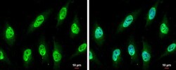
- Immunocytochemistry-Immunofluorescence analysis of NUP62 was performed in HeLa cells fixed in 4% paraformaldehyde at RT for 15 min. Green: NUP62 Polyclonal Antibody (Product # PA5-21882) diluted at 1:750. Blue: Hoechst 33342 staining. Scale bar = 10 µm.

- Submitted by
- Invitrogen Antibodies (provider)
- Main image

- Experimental details
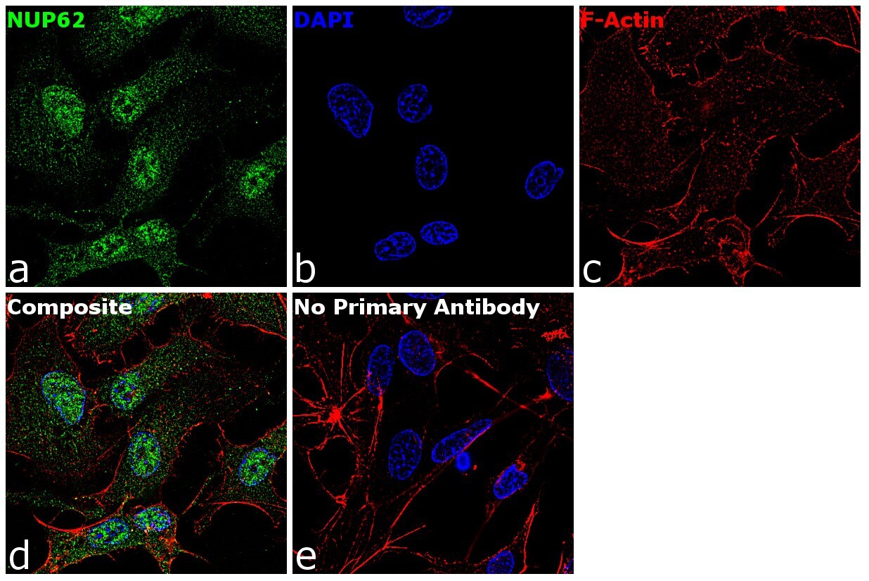
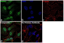
- Immunofluorescence analysis of NUP62 was performed using 70% confluent log phase HepG2 cells. The cells were fixed with 4% paraformaldehyde for 10 minutes, permeabilized with 0.1% Triton™ X-100 for 15 minutes, and blocked with 1% BSA for 1 hour at room temperature. The cells were labeled with NUP62 Rabbit Polyclonal Antibody(Product # PA5-21882) at 5 µg/mL in 0.1% BSA, incubated at 4 degree Celsius overnight and then labeled with Goat anti-Rabbit IgG (H+L) Superclonal™ Secondary Antibody, Alexa Fluor® 488 conjugate (Product # A27034) at a dilution of 1:2000 for 45 minutes at room temperature (Panel a: green). Nuclei (Panel b: blue) were stained with ProLong™ Diamond Antifade Mountant with DAPI (Product # P36962). F-actin (Panel c: red) was stained with Rhodamine Phalloidin (Product # R415, 1:300). Panel d represents the merged image showing Nucleus and cytoplasmic localization. Panel e represents control cells with no primary antibody to assess background. The images were captured at 60X magnification.
- Submitted by
- Invitrogen Antibodies (provider)
- Main image
- Experimental details
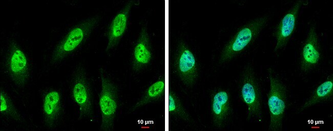
- Immunocytochemistry-Immunofluorescence analysis of NUP62 was performed in HeLa cells fixed in 4% paraformaldehyde at RT for 15 min. Green: NUP62 Polyclonal Antibody (Product # PA5-21882) diluted at 1:750. Blue: Hoechst 33342 staining. Scale bar = 10 µm.
- Submitted by
- Invitrogen Antibodies (provider)
- Main image
- Experimental details
- Immunofluorescence analysis of NUP62 was performed using 70% confluent log phase HepG2 cells. The cells were fixed with 4% paraformaldehyde for 10 minutes, permeabilized with 0.1% Triton™ X-100 for 15 minutes, and blocked with 1% BSA for 1 hour at room temperature. The cells were labeled with NUP62 Rabbit Polyclonal Antibody(Product # PA5-21882) at 5 µg/mL in 0.1% BSA, incubated at 4 degree Celsius overnight and then labeled with Goat anti-Rabbit IgG (Heavy Chain) Superclonal™ Secondary Antibody, Alexa Fluor® 488 conjugate (Product # A27034) at a dilution of 1:2000 for 45 minutes at room temperature (Panel a: green). Nuclei (Panel b: blue) were stained with ProLong™ Diamond Antifade Mountant with DAPI (Product # P36962). F-actin (Panel c: red) was stained with Rhodamine Phalloidin (Product # R415, 1:300). Panel d represents the merged image showing Nucleus and cytoplasmic localization. Panel e represents control cells with no primary antibody to assess background. The images were captured at 60X magnification.

Supportive validation
- Submitted by
- Invitrogen Antibodies (provider)
- Main image

- Experimental details
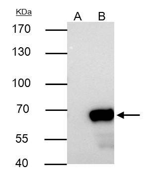
- NUP62 Polyclonal Antibody immunoprecipitates NUP62 protein in IP experiments. IP samples: 293T whole cell extract. A. Control with 4 µg of preimmune Rabbit IgG. B. Immunoprecipitation of NUP62 protein by 4 µg NUP62 Polyclonal Antibody (Product # PA5-21882). 7.5 % SDS-PAGE. The immunoprecipitated NUP62 protein was detected by NUP62 Polyclonal Antibody (Product # PA5-21882) diluted at 1:1,000.

Supportive validation
- Submitted by
- Invitrogen Antibodies (provider)
- Main image
- Experimental details
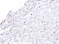
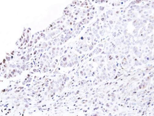
- Immunohistochemical analysis of paraffin-embedded NCIN87 xenograft, using nucleoporin p62 (Product # PA5-21882) antibody at 1:500 dilution. Antigen Retrieval: EDTA based buffer, pH 8.0, 15 min.
- Submitted by
- Invitrogen Antibodies (provider)
- Main image
- Experimental details
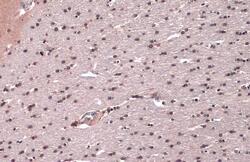
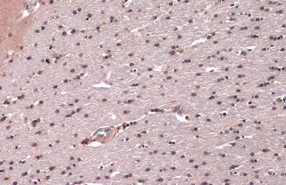
- NUP62 Polyclonal Antibody detects Nucleoporin p62 protein at nucleus by immunohistochemical analysis. Sample: Paraffin-embedded rat brain. Nucleoporin p62 stained by NUP62 Polyclonal Antibody (Product # PA5-21882) diluted at 1:500. Antigen Retrieval: Citrate buffer, pH 6.0, 15 min.
Supportive validation
- Submitted by
- Invitrogen Antibodies (provider)
- Main image

- Experimental details
- NUP62 Polyclonal Antibody immunoprecipitates NUP62 protein in IP experiments. IP samples: 293T whole cell extract. A. Control with 4 µg of preimmune Rabbit IgG. B. Immunoprecipitation of NUP62 protein by 4 µg NUP62 Polyclonal Antibody (Product # PA5-21882). 7.5 % SDS-PAGE. The immunoprecipitated NUP62 protein was detected by NUP62 Polyclonal Antibody (Product # PA5-21882) diluted at 1:1,000.