Explore
 Validate
 Learn
Learn Western blot
 Immunocytochemistry
Antibody data
- Antibody Data
- Antigen structure
- References [3]
- Comments [0]
- Validations
- Immunocytochemistry [7]
- Other assay [4]
Submit
Validation data
Reference
Comment
Report error
- Product number
- MA1-10031 - Provider product page

- Provider
- Invitrogen Antibodies
- Product name
- NUP107 Monoclonal Antibody (39C7)
- Antibody type
- Monoclonal
- Antigen
- Other
- Description
- For immunofluorescence on yeast cells, try MCA-39C7 diluted 1:100 to 1:500. For immunofluorescence on mammalian cells try at 1:50 to 1:100.
- Reactivity
- Human, Mouse, Rat, Bovine, Chicken/Avian, Drosophila, Porcine, Yeast
- Host
- Mouse
- Isotype
- IgG
- Antibody clone number
- 39C7
- Vial size
- 500 μL
- Concentration
- Conc. Not Determined
- Storage
- Store at 4°C short term. For long term storage, store at -20°C, avoiding freeze/thaw cycles.
Submitted references Stimulated emission depletion microscopy with a single depletion laser using five fluorochromes and fluorescence lifetime phasor separation.
Nuclear Pore Complexes Cluster in Dysmorphic Nuclei of Normal and Progeria Cells during Replicative Senescence.
G(4)C(2) Repeat RNA Initiates a POM121-Mediated Reduction in Specific Nucleoporins in C9orf72 ALS/FTD.
Gonzalez Pisfil M, Nadelson I, Bergner B, Rottmeier S, Thomae AW, Dietzel S
Scientific reports 2022 Aug 18;12(1):14027
Scientific reports 2022 Aug 18;12(1):14027
Nuclear Pore Complexes Cluster in Dysmorphic Nuclei of Normal and Progeria Cells during Replicative Senescence.
Röhrl JM, Arnold R, Djabali K
Cells 2021 Jan 14;10(1)
Cells 2021 Jan 14;10(1)
G(4)C(2) Repeat RNA Initiates a POM121-Mediated Reduction in Specific Nucleoporins in C9orf72 ALS/FTD.
Coyne AN, Zaepfel BL, Hayes L, Fitchman B, Salzberg Y, Luo EC, Bowen K, Trost H, Aigner S, Rigo F, Yeo GW, Harel A, Svendsen CN, Sareen D, Rothstein JD
Neuron 2020 Sep 23;107(6):1124-1140.e11
Neuron 2020 Sep 23;107(6):1124-1140.e11
No comments: Submit comment
Supportive validation
- Submitted by
- Invitrogen Antibodies (provider)
- Main image
- Experimental details
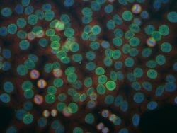
- Immunohistochemistry analysis of human HeLa cells stained with monoclonal antibody, Product # MA1-10031, which binds to a nuclear pore complex antigen.

- Submitted by
- Invitrogen Antibodies (provider)
- Main image

- Experimental details
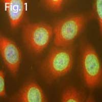
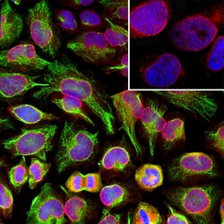
- Immunofluorescent analysis of NUP107 using a monoclonal antibody (Product # MA1-10031).
- Submitted by
- Invitrogen Antibodies (provider)
- Main image
- Experimental details
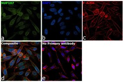
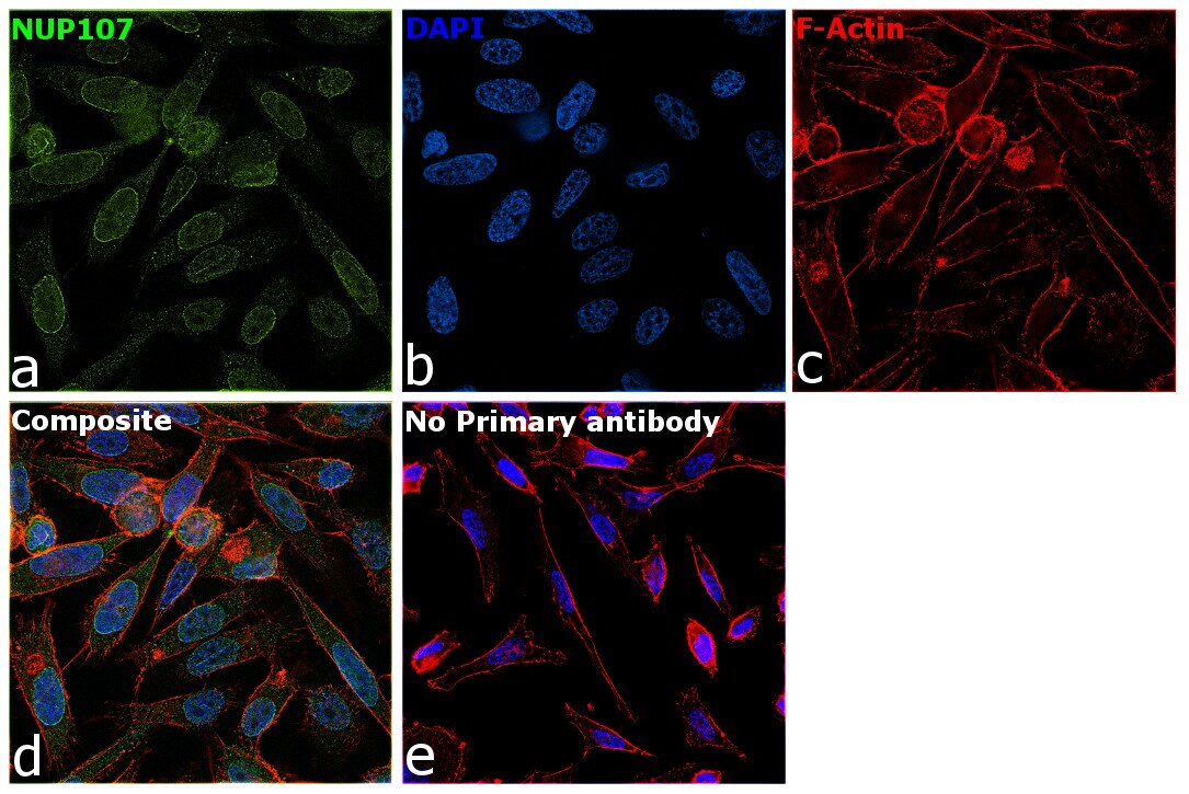
- Immunofluorescence analysis of NUP107 was performed using 70% confluent log phase PC-3 cells. The cells were fixed with 4% Paraformaldehyde for 10 minutes, permeabilized with 0.1% Triton™ X-100 for 10 minutes, and blocked with 2% BSA for 10 minutes at room temperature. The cells were labeled with NUP107 Monoclonal Antibody (39C7) (Product # MA1-10031) at 1:100 dilution in 0.1% BSA, incubated at 4 degree celsius overnight and then labeled with Goat anti-Mouse IgG (H+L) Highly Cross-Adsorbed Secondary Antibody, Alexa Fluor Plus 488 (Product # A32723, 1:2000 dilution) for 45 minutes at room temperature (Panel a: Green). Nuclei (Panel b: Blue) were stained with SlowFade® Gold Antifade Mountant with DAPI (Product # S36938). F-actin (Panel c: Red) was stained with Rhodamine Phalloidin (Product # R415, 1:300). Panel d represents the merged image showing nuclear membrane localization. Panel e represents control cells with no primary antibody to assess background. The images were captured at 60X magnification.

- Submitted by
- Invitrogen Antibodies (provider)
- Main image
- Experimental details
- Immunofluorescent analysis of NUP107 in HeLa cells. Cells were stained red using a NUP107 monoclonal antibody (Product # MA1-10031) at a dilution of 1:1,000, stained green with a Vimentin polyclonal antibody (Product # PA1-10003) at a dilution of 1:10,000, and nuclear DNA was stained blue with DAPI.

- Submitted by
- Invitrogen Antibodies (provider)
- Main image

- Experimental details
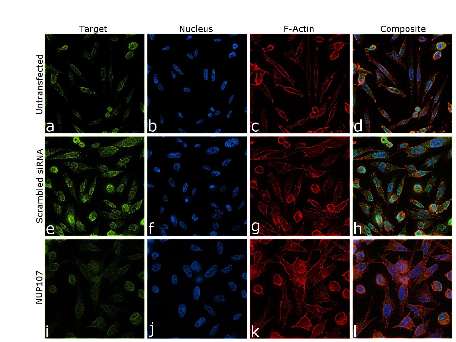
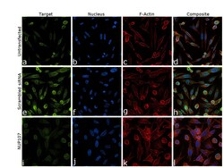
- Knockdown of NUP107 was achieved by transfecting PC-3 cells with NUP107 specific siRNA (Silencer® select Product # s32728, s32727). Immunofluorescence analysis was performed on untransfected PC-3 cells (panel a,d), transfected with non-specific scrambled siRNA (panels b,e) and transfected with NUP107 specific siRNA (panel c,f). Cells were fixed, permeabilized, and labelled with NUP107 Monoclonal Antibody (39C7) (Product # MA1-10031), (1:100 dilution) followed by Goat anti-Mouse IgG (H+L) Highly Cross-Adsorbed Secondary Antibody, Alexa Fluor Plus 488 (Product # A32723, 1:2000 dilution). Nuclei (blue) were stained with SlowFade® Gold Antifade Mountant with DAPI (Product # S36938), and Rhodamine Phalloidin (Product # R415, 1:300) was used for cytoskeletal F-actin (Red) staining. Partial reduction of specific signal was observed upon siRNA mediated knockdown (panel c,f) confirming specificity of the antibody to NUP107 (Green). The Images were captured at 60X magnification.
- Submitted by
- Invitrogen Antibodies (provider)
- Main image

- Experimental details
- Immunofluorescence analysis of NUP107 was performed using 70% confluent log phase PC-3 cells. The cells were fixed with 4% Paraformaldehyde for 10 minutes, permeabilized with 0.1% Triton™ X-100 for 10 minutes, and blocked with 2% BSA for 10 minutes at room temperature. The cells were labeled with NUP107 Monoclonal Antibody (39C7) (Product # MA1-10031) at 1:100 dilution in 0.1% BSA, incubated at 4 degree celsius overnight and then labeled with Goat anti-Mouse IgG (H+L) Highly Cross-Adsorbed Secondary Antibody, Alexa Fluor Plus 488 (Product # A32723, 1:2000 dilution) for 45 minutes at room temperature (Panel a: Green). Nuclei (Panel b: Blue) were stained with SlowFade® Gold Antifade Mountant with DAPI (Product # S36938). F-actin (Panel c: Red) was stained with Rhodamine Phalloidin (Product # R415, 1:300). Panel d represents the merged image showing nuclear membrane localization. Panel e represents control cells with no primary antibody to assess background. The images were captured at 60X magnification.
- Submitted by
- Invitrogen Antibodies (provider)
- Main image
- Experimental details
- Knockdown of NUP107 was achieved by transfecting PC-3 cells with NUP107 specific siRNA (Silencer® select Product # s32728, s32727). Immunofluorescence analysis was performed on untransfected PC-3 cells (panel a,d), transfected with non-specific scrambled siRNA (panels b,e) and transfected with NUP107 specific siRNA (panel c,f). Cells were fixed, permeabilized, and labelled with NUP107 Monoclonal Antibody (39C7) (Product # MA1-10031), (1:100 dilution) followed by Goat anti-Mouse IgG (H+L) Highly Cross-Adsorbed Secondary Antibody, Alexa Fluor Plus 488 (Product # A32723, 1:2000 dilution). Nuclei (blue) were stained with SlowFade® Gold Antifade Mountant with DAPI (Product # S36938), and Rhodamine Phalloidin (Product # R415, 1:300) was used for cytoskeletal F-actin (Red) staining. Partial reduction of specific signal was observed upon siRNA mediated knockdown (panel c,f) confirming specificity of the antibody to NUP107 (Green). The Images were captured at 60X magnification.

Supportive validation
- Submitted by
- Invitrogen Antibodies (provider)
- Main image

- Experimental details
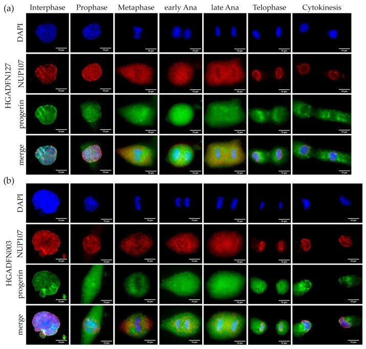
- Figure 2 Progerin aggregates did not colocalize with NUP107 in mitotic Hutchinson-Gilford progeria syndrome (HGPS) cells. Immunocytochemistry of ( a ) HGADFN127 and ( b ) HGADFN003 fibroblasts using alphai-NUP107 and alpha-progerin antibody, counterstained with DAPI. In metaphase to cytokinesis, progerin forms aggregates in the cytoplasm of mitotic HGPS cells, which are absent in control cells. Recruitment of NUP107 to late anaphase/telophase chromosomes is not delayed in HGPS cells. n >= 3.
- Submitted by
- Invitrogen Antibodies (provider)
- Main image
- Experimental details
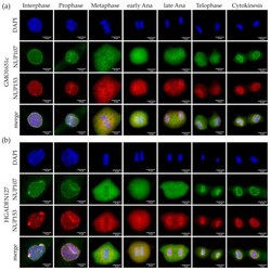
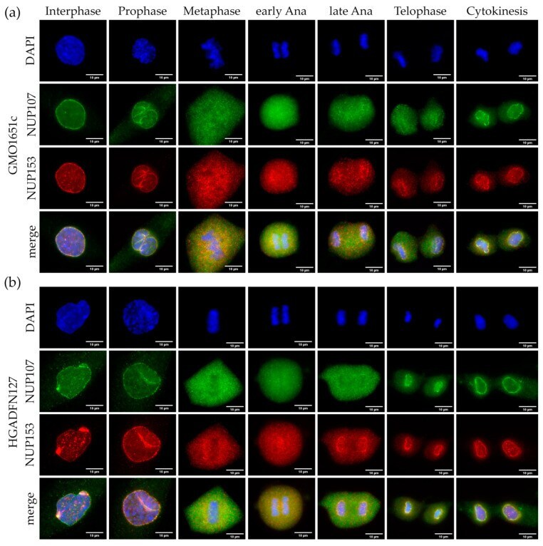
- Figure 4 Recruitment of basket nucleoporin NUP153 and scaffold nucleoporin NUP107 was not affected in HGPS. Immunocytochemistry of ( a ) control (GMO1651c) and ( b ) HGPS (HGADFN127) fibroblasts using alpha-NUP107 and alpha-NUP153 antibodies, counterstained with DAPI. NPCs cluster in interphase HGPS cells ( b ), unlike the evenly distributed punctate pattern in control interphase ( a ). Recruitment of NUP153 to anaphase chromosomes is not affected in HGPS cells when compared to control. No defect in NUP153 or NUP107 localization can be observed in HGPS from prophase to cytokinesis. n >= 3.
- Submitted by
- Invitrogen Antibodies (provider)
- Main image
- Experimental details
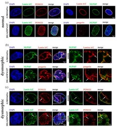
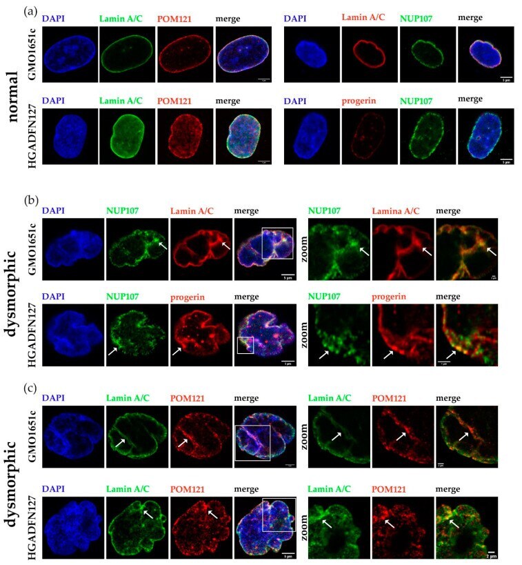
- Figure 9 NPCs clustered in folds of dysmorphic nuclear envelopes and co-localized with progerin/Lamin A aggregates trapped at the nuclear membrane. ( a ) Representative images of normal control (GMO1651c) and HGPS (HGADFN127) nuclei stained with alpha-NUP107/POM121/LA/progerin antibodies, counterstained with DAPI. ( b ) Representative images of dysmorphic control and HGPS nuclei, stained with alpha-NUP107 and alpha-LA/C or progerin antibodies, counterstained with DAPI. ( c ) Representative images of dysmorphic control and HGPS nuclei, stained with alpha-POM121 and LA antibodies, counterstained with DAPI. Arrows indicate regions with clustered NPCs, outlines indicate zoomed in section of nuclei. All images are taken from z-stack and are provided as X-Z view. Zoom factor: ( b ) GMO1651c 15x, HGADFN127 25x; ( c ) GMO1651c 9x, HGADFN127 12x.
- Submitted by
- Invitrogen Antibodies (provider)
- Main image
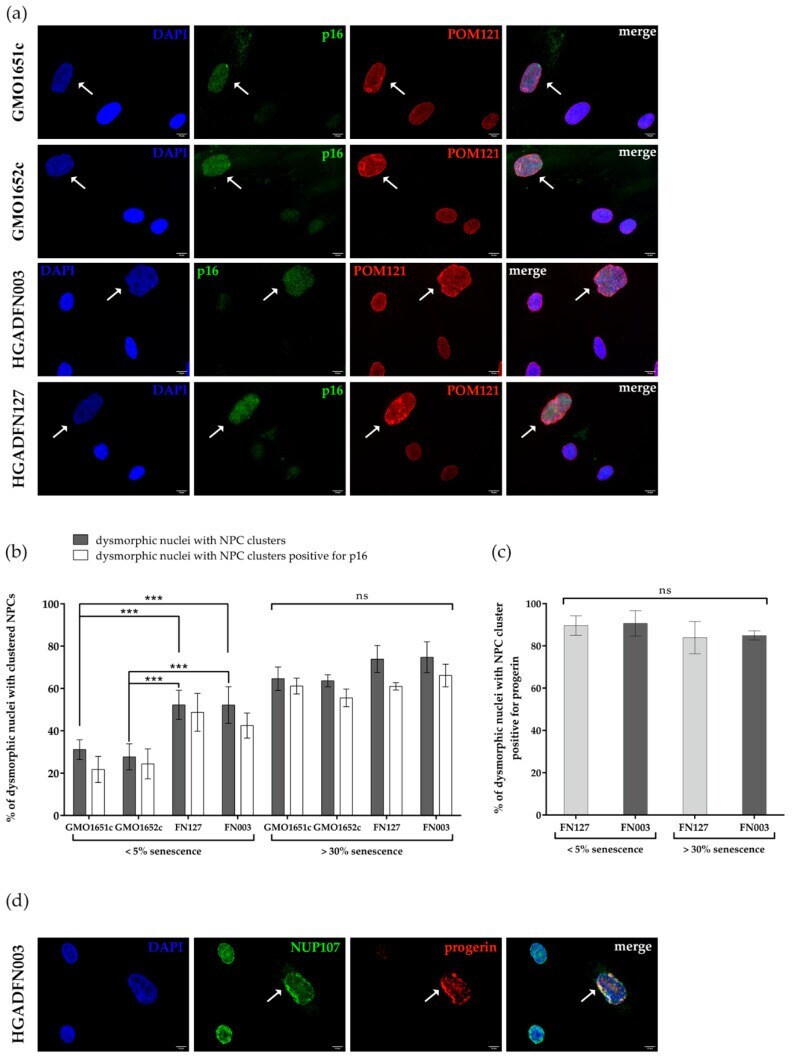
- Experimental details
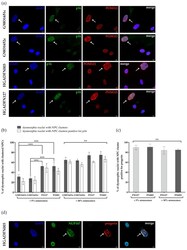
- Figure 10 Number of dysmorphic nuclei with clustered NPCs increased with replicative senescence. ( a ) Representative images of cells stained with alpha-p16 and alpha-POM121 antibodies, counterstained with DAPI. White arrows indicate dysmorphic nuclei with clustered NPCs and elevated p16. ( b ) The numbers of dysmorphic nuclei with clustered NPCs, in cultures =30% senescence of control and HGPS cells, were determined by counting dysmorphic nuclei positive for clustered NPCs and p16 positive signal, counterstained with DAPI. Differences between young and old cells in each cell line are significant. ( c ) The numbers of dysmorphic nuclei with clustered NPCs and progerin positive. On average, 87% of dysmorphic HGPS nuclei with clustered NPCs are positive for progerin. No significant difference could be determined depending on replicative senescence. ( d ) Representative images of HGADFN003 stained with alpha-NUP107 and alpha-progerin antibodies, counterstained with DAPI. White arrows indicate NUP107 clusters overlapping with strong progerin signal. At 5% senescence, HGPS cell passages were =P21. Values are presented as mean +- SD (n >= 3), ns p > 0.05, *** p < 001, ( b ) two-way ANOVA with Tukey's multiple comparison test, ( c ) one-way ANOVA with Sidak's multiple comparison test.