 Explore
 Validate
 Learn
Learn Western blot
 Immunocytochemistry
 Immunoprecipitation
Antibody data
- Antibody Data
- Antigen structure
- References [1]
- Comments [0]
- Validations
- Immunocytochemistry [9]
- Immunohistochemistry [16]
- Other assay [1]
Submit
Validation data
Reference
Comment
Report error
- Product number
- PA1-32451 - Provider product page

- Provider
- Invitrogen Antibodies
- Product name
- NuMA Polyclonal Antibody
- Antibody type
- Polyclonal
- Antigen
- Purifed from natural sources
- Description
- Store product as a concentrated solution. Centrifuge briefly prior to opening the vial.
- Reactivity
- Human, Mouse, Rat
- Host
- Rabbit
- Isotype
- IgG
- Vial size
- 100 μL
- Concentration
- Conc. Not Determined
- Storage
- Store at 4°C short term. For long term storage, store at -20°C, avoiding freeze/thaw cycles.
Submitted references Neuronal life or death linked to depression treatment: the interplay between drugs and their stress-related outcomes relate to single or combined drug therapies.
Solek P, Koszla O, Mytych J, Badura J, Chelminiak Z, Cuprys M, Fraczek J, Tabecka-Lonczynska A, Koziorowski M
Apoptosis : an international journal on programmed cell death 2019 Oct;24(9-10):773-784
Apoptosis : an international journal on programmed cell death 2019 Oct;24(9-10):773-784
No comments: Submit comment
Supportive validation
- Submitted by
- Invitrogen Antibodies (provider)
- Main image

- Experimental details
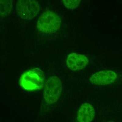
- Immunocytochemistry-Immunofluorescence analysis of NuMA in HeLa cells. Cells were fixed with 3.5% formaldehyde and NuMA Polyclonal Antibody (Product # PA1-32451) at a dilution of 1:1000 was used as a primary antibody. The staining pattern shows the typical pattern for NuMA in the cell nucleus during interphase and on spindles during mitosis.
- Submitted by
- Invitrogen Antibodies (provider)
- Main image

- Experimental details
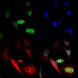
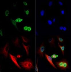
- Immunocytochemistry-Immunofluorescence analysis of NuMA in HeLa cells using NuMA Polyclonal Antibody (Product # PA1-32451) (Green). Red : Tubulin. Blue: DAPI.

- Submitted by
- Invitrogen Antibodies (provider)
- Main image
- Experimental details
- Immunocytochemistry-Immunofluorescence analysis of NuMA in HeLa cells using NuMA Polyclonal Antibody (Product # PA1-32451) (Green). Red : Tubulin. Blue: DAPI.

- Submitted by
- Invitrogen Antibodies (provider)
- Main image

- Experimental details
- Immunocytochemistry-Immunofluorescence analysis of NuMA in HeLa cells. Cells were fixed with 3.5% formaldehyde and NuMA Polyclonal Antibody (Product # PA1-32451) at a dilution of 1:1000 was used as a primary antibody. The staining pattern shows the typical pattern for NuMA in the cell nucleus during interphase and on spindles during mitosis.

- Submitted by
- Invitrogen Antibodies (provider)
- Main image
- Experimental details
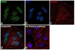
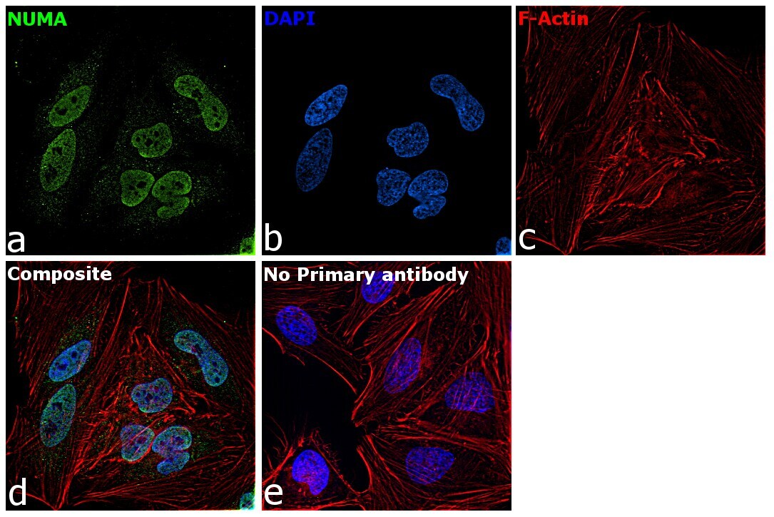
- Immunofluorescence analysis of NuMA was performed using HeLa cells. The cells were fixed with 4% paraformaldehyde for 10 minutes, permeabilized with 0.1% Triton™ X-100 for 15 minutes, and blocked with 2% BSA for 1 hour at room temperature. The cells were labeled with NuMA Polyclonal Antibody (Product # PA1-32451) at 1:200 dilution in 0.1% BSA, incubated at 4 degree celsius overnight and then labeled with Goat anti-Rabbit IgG (H+L) Superclonal™ Recombinant Secondary Antibody, Alexa Fluor® 488 conjugate (Product # A27034) at a dilution of 1:2000 for 45 minutes at room temperature (Panel a: green). Nuclei (Panel b: blue) were stained with ProLong™ Diamond Antifade Mountant with DAPI (Product # P36962). F-actin (Panel c: red) was stained with Rhodamine Phalloidin (Product # R415, 1:300). Panel d represents the merged image showing nuclear localization. Panel e represents control cells with no primary antibody to assess background. The images were captured at 60X magnification.

- Submitted by
- Invitrogen Antibodies (provider)
- Main image

- Experimental details
- Immunocytochemistry-Immunofluorescence analysis of NuMA in HeLa cells. Cells were fixed with 3.5% formaldehyde and NuMA Polyclonal Antibody (Product # PA1-32451) at a dilution of 1:1000 was used as a primary antibody. The staining pattern shows the typical pattern for NuMA in the cell nucleus during interphase and on spindles during mitosis.

- Submitted by
- Invitrogen Antibodies (provider)
- Main image

- Experimental details
- Immunocytochemistry-Immunofluorescence analysis of NuMA in HeLa cells using NuMA Polyclonal Antibody (Product # PA1-32451) (Green). Red : Tubulin. Blue: DAPI.
- Submitted by
- Invitrogen Antibodies (provider)
- Main image
- Experimental details
- Immunocytochemistry-Immunofluorescence analysis of NuMA in HeLa cells using NuMA Polyclonal Antibody (Product # PA1-32451) (Green). Red : Tubulin. Blue: DAPI.

- Submitted by
- Invitrogen Antibodies (provider)
- Main image

- Experimental details
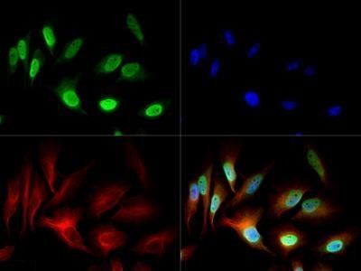
- Immunofluorescence analysis of NuMA was performed using HeLa cells. The cells were fixed with 4% paraformaldehyde for 10 minutes, permeabilized with 0.1% Triton™ X-100 for 15 minutes, and blocked with 2% BSA for 1 hour at room temperature. The cells were labeled with NuMA Polyclonal Antibody (Product # PA1-32451) at 1:200 dilution in 0.1% BSA, incubated at 4 degree celsius overnight and then labeled with Goat anti-Rabbit IgG (Heavy Chain) Superclonal™ Recombinant Secondary Antibody, Alexa Fluor® 488 conjugate (Product # A27034) at a dilution of 1:2000 for 45 minutes at room temperature (Panel a: green). Nuclei (Panel b: blue) were stained with ProLong™ Diamond Antifade Mountant with DAPI (Product # P36962). F-actin (Panel c: red) was stained with Rhodamine Phalloidin (Product # R415, 1:300). Panel d represents the merged image showing nuclear localization. Panel e represents control cells with no primary antibody to assess background. The images were captured at 60X magnification.
Supportive validation
- Submitted by
- Invitrogen Antibodies (provider)
- Main image
- Experimental details
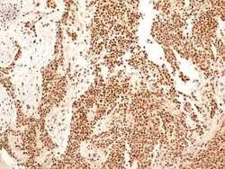
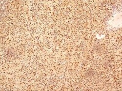
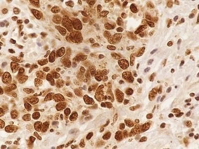
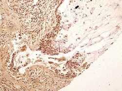
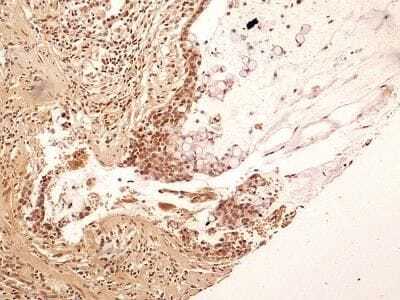
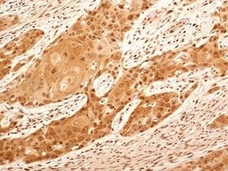
- Immunohistochemistry (Paraffin) analysis of NuMA in human bladder cancer tissue using NuMA Polyclonal Antibody (Product # PA1-32451) at a dilution of 5 µg/mL.
- Submitted by
- Invitrogen Antibodies (provider)
- Main image

- Experimental details
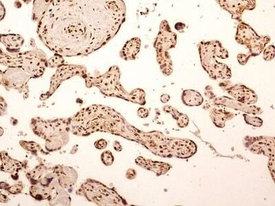
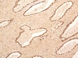
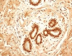
- Immunohistochemistry (Paraffin) analysis of NuMA in human placenta tissue using NuMA Polyclonal Antibody (Product # PA1-32451) at a dilution of 5 µg/mL.
- Submitted by
- Invitrogen Antibodies (provider)
- Main image

- Experimental details
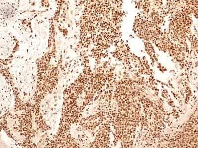
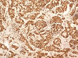
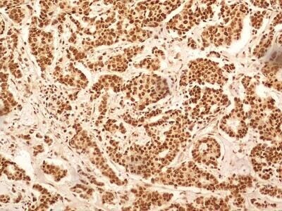
- Immunohistochemistry (Paraffin) analysis of NuMA in poorly differentiated human endometrial carcinoma tissue using NuMA Polyclonal Antibody (Product # PA1-32451) at a dilution of 5 µg/mL.
- Submitted by
- Invitrogen Antibodies (provider)
- Main image
- Experimental details
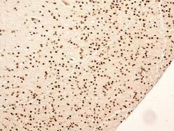
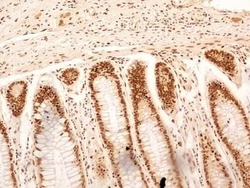
- Immunohistochemistry (Paraffin) analysis of NuMA in human brain tissue using NuMA Polyclonal Antibody (Product # PA1-32451) at a dilution of 5 µg/mL.

- Submitted by
- Invitrogen Antibodies (provider)
- Main image
- Experimental details
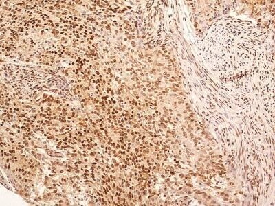
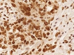
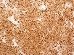
- Immunohistochemistry (Paraffin) analysis of NuMA in human breast cancer tissue using NuMA Polyclonal Antibody (Product # PA1-32451) at a dilution of 5 µg/mL.
- Submitted by
- Invitrogen Antibodies (provider)
- Main image
- Experimental details
- Immunohistochemistry (Paraffin) analysis of NuMA in human spleen tissue using NuMA Polyclonal Antibody (Product # PA1-32451) at a dilution of 5 µg/mL.

- Submitted by
- Invitrogen Antibodies (provider)
- Main image
- Experimental details
- Immunohistochemistry (Paraffin) analysis of NuMA in human rectal adenocarcinoma tissue using NuMA Polyclonal Antibody (Product # PA1-32451) at a dilution of 5 µg/mL.

- Submitted by
- Invitrogen Antibodies (provider)
- Main image
- Experimental details
- Immunohistochemistry (Paraffin) analysis of NuMA in human pulmonary squamous cell carcinoma tissue using NuMA Polyclonal Antibody (Product # PA1-32451) at a dilution of 5 µg/mL.
- Submitted by
- Invitrogen Antibodies (provider)
- Main image
- Experimental details
- Immunohistochemistry (Paraffin) analysis of NuMA in human stomach adenocarcinoma tissue using NuMA Polyclonal Antibody (Product # PA1-32451) at a dilution of 5 µg/mL.
- Submitted by
- Invitrogen Antibodies (provider)
- Main image
- Experimental details
- Immunohistochemistry (Paraffin) analysis of NuMA in human esophageal squamous cell carcinoma (SCC) tissue using NuMA Polyclonal Antibody (Product # PA1-32451) at a dilution of 5 µg/mL.

- Submitted by
- Invitrogen Antibodies (provider)
- Main image
- Experimental details
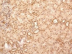
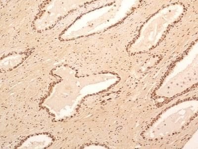
- Immunohistochemistry (Paraffin) analysis of NuMA in human kidney tissue using NuMA Polyclonal Antibody (Product # PA1-32451) at a dilution of 5 µg/mL.

- Submitted by
- Invitrogen Antibodies (provider)
- Main image

- Experimental details
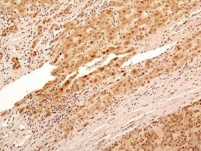
- Immunohistochemistry (Paraffin) analysis of NuMA in human liver cancer tissue using NuMA Polyclonal Antibody (Product # PA1-32451) at a dilution of 5 µg/mL.
- Submitted by
- Invitrogen Antibodies (provider)
- Main image
- Experimental details
- Immunohistochemistry (Paraffin) analysis of NuMA in human prostate tissue using NuMA Polyclonal Antibody (Product # PA1-32451) at a dilution of 5 µg/mL.
- Submitted by
- Invitrogen Antibodies (provider)
- Main image
- Experimental details
- Immunohistochemistry (Paraffin) analysis of NuMA in small bowel stromal cancer tissue using NuMA Polyclonal Antibody (Product # PA1-32451) at a dilution of 5 µg/mL.

- Submitted by
- Invitrogen Antibodies (provider)
- Main image
- Experimental details
- Immunohistochemistry (Paraffin) analysis of NuMA in human breast tissue using NuMA Polyclonal Antibody (Product # PA1-32451) at a dilution of 5 µg/mL.

- Submitted by
- Invitrogen Antibodies (provider)
- Main image
- Experimental details
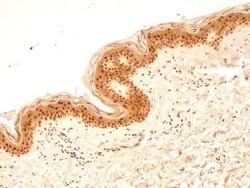
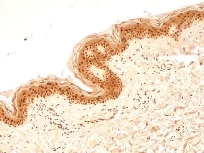
- Immunohistochemistry (Paraffin) analysis of NuMA in human skin tissue using NuMA Polyclonal Antibody (Product # PA1-32451) at a dilution of 5 µg/mL.
Supportive validation
- Submitted by
- Invitrogen Antibodies (provider)
- Main image
- Experimental details
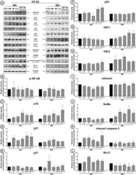
- Fig. 4 Antidepressants-mediated effect on cellular protein content. HT-22 cells were treated with antidepressants for 48 and 96 h and densitometry analysis of NF-kappaB ( b ), p16 ( c ), p21 ( d ), p27 ( e ), p53 ( f ), TRF1 ( g ), TRF2 ( h ), calnexin ( i ), NuMa ( j ), cleaved caspase 3 ( k ), Bcl-2 ( l ) was evaluated. Representative Western Blots are presented ( a ). Bars indicate SD, n = 3, *** /^^^ p < 0.001, ** /^^ p < 0.01, * /^ p < 0.05, no indication--no statistical significance (one-way ANOVA and Dunnett's a posteriori test)
