 Explore
 Validate
 Learn
Learn Western blot
 Immunohistochemistry
Antibody data
- Antibody Data
- Antigen structure
- References [1]
- Comments [0]
- Validations
- Immunohistochemistry [1]
Submit
Validation data
Reference
Comment
Report error
- Product number
- HPA018843 - Provider product page

- Provider
- Atlas Antibodies
- Proper citation
- Atlas Antibodies Cat#HPA018843, RRID:AB_2669948
- Product name
- Anti-CAPG
- Antibody type
- Polyclonal
- Description
- Polyclonal Antibody against Human CAPG, Gene description: capping protein (actin filament), gelsolin-like, Alternative Gene Names: AFCP, MCP, Validated applications: WB, IHC, Uniprot ID: P40121, Storage: Store at +4°C for short term storage. Long time storage is recommended at -20°C.
- Reactivity
- Human, Mouse, Rat
- Host
- Rabbit
- Conjugate
- Unconjugated
- Isotype
- IgG
- Vial size
- 100 µl
- Concentration
- 0.05 mg/ml
- Storage
- Store at +4°C for short term storage. Long time storage is recommended at -20°C.
- Handling
- The antibody solution should be gently mixed before use.
Submitted references A High-throughput Bead-based Affinity Assay Enables Analysis of Genital Protein Signatures in Women At Risk of HIV Infection
Månberg A, Bradley F, Qundos U, Guthrie B, Birse K, Noël-Romas L, Lindskog C, Bosire R, Kiarie J, Farquhar C, Burgener A, Nilsson P, Broliden K
Molecular & Cellular Proteomics 2019;18(3):461-476
Molecular & Cellular Proteomics 2019;18(3):461-476
No comments: Submit comment
Supportive validation
- Submitted by
- Atlas Antibodies (provider)
- Enhanced method
- Orthogonal validation
- Main image
- Experimental details
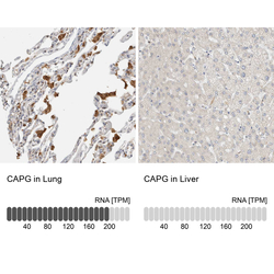
- Immunohistochemistry analysis in human lung and liver tissues using HPA018843 antibody. Corresponding CAPG RNA-seq data are presented for the same tissues.
- Sample type
- Human
- Protocol
- Protocol
