Explore
 Validate
 Learn
Learn Western blot
Antibody data
- Antibody Data
- Antigen structure
- References [1]
- Comments [0]
- Validations
- Western blot [1]
Submit
Validation data
Reference
Comment
Report error
- Product number
- HPA019092 - Provider product page

- Provider
- Atlas Antibodies
- Proper citation
- Atlas Antibodies Cat#HPA019092, RRID:AB_1845980
- Product name
- Anti-CAPG
- Antibody type
- Polyclonal
- Description
- Polyclonal Antibody against Human CAPG, Gene description: capping protein (actin filament), gelsolin-like, Alternative Gene Names: AFCP, MCP, Validated applications: IHC, WB, Uniprot ID: P40121, Storage: Store at +4°C for short term storage. Long time storage is recommended at -20°C.
- Reactivity
- Human
- Host
- Rabbit
- Conjugate
- Unconjugated
- Isotype
- IgG
- Vial size
- 100 µl
- Concentration
- 0.05 mg/ml
- Storage
- Store at +4°C for short term storage. Long time storage is recommended at -20°C.
- Handling
- The antibody solution should be gently mixed before use.
Submitted references A High-throughput Bead-based Affinity Assay Enables Analysis of Genital Protein Signatures in Women At Risk of HIV Infection
Månberg A, Bradley F, Qundos U, Guthrie B, Birse K, Noël-Romas L, Lindskog C, Bosire R, Kiarie J, Farquhar C, Burgener A, Nilsson P, Broliden K
Molecular & Cellular Proteomics 2019;18(3):461-476
Molecular & Cellular Proteomics 2019;18(3):461-476
No comments: Submit comment
Enhanced validation
- Submitted by
- 57e50c6745390
- Enhanced method
- Orthogonal validation
- Main image

- Experimental details
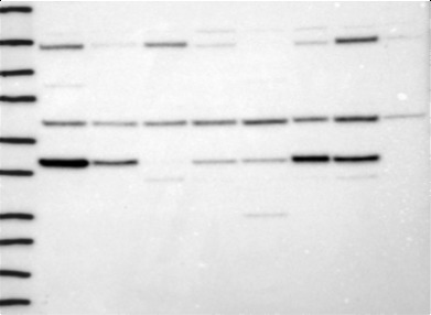
- Orthogonal validation by targeted mass spectrometry with stable isotope labeled antigen (QPrEST). Target protein was quantified across eight cell lines and the same cell lysate was subjected for WB-analysis.
- Sample type
- Cell lysates
- Validation comment
- Multiple western blot bands detected. One of expected molecular weight shows excellent correlation if compared to quantitative data determined by MS.
- Primary Ab dilution
- 0.2 ug/ml
- Other comments
- Multiple western blot bands detected. Band of expected molecular weight shows excellent correlation if compared to quantitative data determined by MS. This is thereby considered as validated for Western Blot applications, with additional cross-reactive band detected.
- Conjugate
- Horseradish Peroxidase
- Secondary Ab
- Secondary Ab
- Secondary Ab dilution
- 1:4000
- Protocol
- Protocol
- Correlation
- 0.97
- p-value
- 0.001
- Number of samples
- 8