Explore
 Validate
 Learn
LearnGTX129433
antibody from GeneTex
Targeting: ARID1A
B120, BAF250, BAF250a, C10rf4, C1orf4, P270, SMARCF1
 Western blot
Antibody data
- Antibody Data
- Antigen structure
- References [0]
- Comments [0]
- Validations
- Western blot [7]
- Immunocytochemistry [1]
- Immunoprecipitation [1]
- Immunohistochemistry [2]
Submit
Validation data
Reference
Comment
Report error
- Product number
- GTX129433 - Provider product page

- Provider
- GeneTex
- Product name
- ARID1A antibody
- Antibody type
- Polyclonal
- Reactivity
- Human
- Host
- Rabbit
No comments: Submit comment
Supportive validation
- Submitted by
- GeneTex (provider)
- Main image
- Experimental details
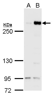
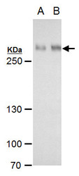
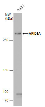
- ARID1A antibody detects ARID1A protein by Western blot analysis.A. 30 µg 293T B. 30 µg of human ARID1A-transfected 293T cells5 % SDS-PAGEARID1A antibody (GTX129433) dilution: 1:5000
- Validation comment
- WB
- Submitted by
- GeneTex (provider)
- Main image
- Experimental details
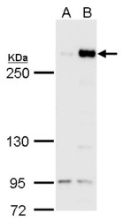
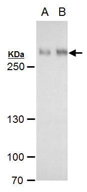
- ARID1A antibody detects ARID1A protein by Western blot analysis.A. 30 µg Huh7 B. 30 µg Hep3B C. 30 µg HepG2 5 % SDS-PAGEARID1A antibody (GTX129433) dilution: 1:1000
- Validation comment
- WB

- Submitted by
- GeneTex (provider)
- Main image
- Experimental details
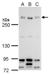
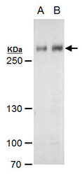
- ARID1A antibody detects ARID1A protein by western blot analysis.A. 30 £gg HepG2 whole cell extractB. 30 £gg HepG2 nuclear extract5 % SDS-PAGEARID1A antibody (GTX129433) dilution: 1:1000
- Submitted by
- GeneTex (provider)
- Main image
- Experimental details
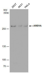
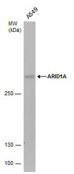
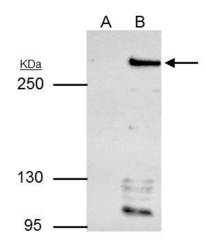
- ARID1A antibody detects ARID1A protein by western blot analysis. Various whole cell extracts (30 £gg) were separated by 5% SDS-PAGE, and the membrane was blotted with ARID1A antibody (GTX129433) diluted by 1:1000.

- Submitted by
- GeneTex (provider)
- Main image
- Experimental details
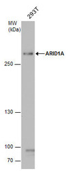
- ARID1A antibody detects ARID1A protein by western blot analysis. Whole cell extracts (30 £gg) was separated by 5% SDS-PAGE, and the membrane was blotted with ARID1A antibody (GTX129433) diluted by 1:1000.
- Submitted by
- GeneTex (provider)
- Main image
- Experimental details
- ARID1A antibody detects ARID1A protein by western blot analysis. Whole cell extracts (30 £gg) was separated by 5% SDS-PAGE, and the membrane was blotted with ARID1A antibody (GTX129433) diluted by 1:1000.

- Submitted by
- GeneTex (provider)
- Main image
- Experimental details
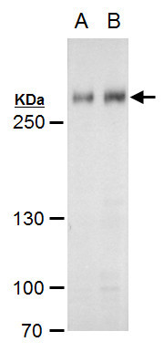
- ARID1A antibody detects ARID1A protein by Western blot analysis.A. 30 £gg HepG2 whole cell extractB. 30 £gg HepG2 nuclear extract5 % SDS-PAGEARID1A antibody (GTX129433) dilution: 1:1000
Supportive validation
- Submitted by
- GeneTex (provider)
- Main image
- Experimental details
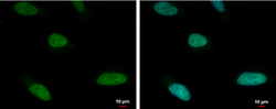
- ARID1A antibody detects ARID1A protein at nucleus by immunofluorescent analysis.Sample: HeLa cells were fixed in 4% paraformaldehyde at RT for 15 min.Green: ARID1A protein stained by ARID1A antibody (GTX129433) diluted at 1:500.Blue: Hoechst 33342 staining.

Supportive validation
- Submitted by
- GeneTex (provider)
- Main image
- Experimental details
- ARID1A antibody immunoprecipitates ARID1A protein in IP experiments.IP samples: HepG2 whole cell extractA. Control with 4 £gg of preimmune Rabbit IgGB. Immunoprecipitation of ARID1A protein by 4 £gg ARID1A antibody (GTX129433)5 % SDS-PAGEThe immunoprecipitated ARID1A protein was detected by ARID1A antibody (GTX129433) diluted at 1:500.[EasyBlot anti-rabbit IgG (GTX221666-01) was used as a secondary reagent]

Supportive validation
- Submitted by
- GeneTex (provider)
- Main image
- Experimental details
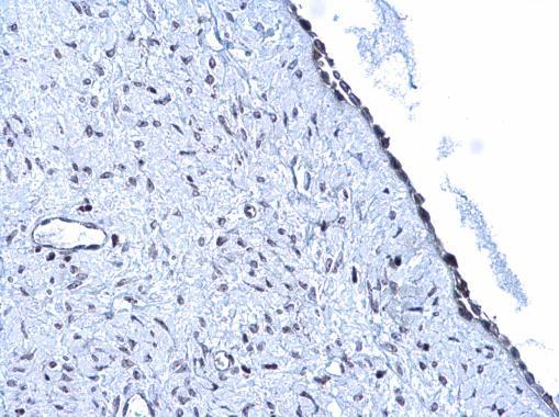
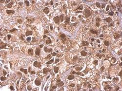
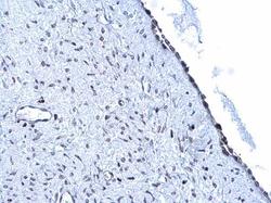
- ARID1A antibody detects ARID1A protein at nucleus on human cervical carcinoma by immunohistochemical analysis. Sample: Paraffin-embedded human cervical carcinoma. ARID1A antibody (GTX129433) dilution: 1:500.
- Submitted by
- GeneTex (provider)
- Main image
- Experimental details
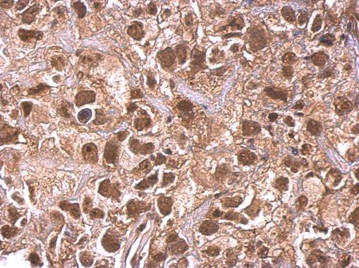
- ARID1A antibody detects ARID1A protein at nucleus on human endometrial carcinoma by immunohistochemical analysis. Sample: Paraffin-embedded human endometrial carcinoma. ARID1A antibody (GTX129433) diluted at 1:500.