Explore
 Validate
 Learn
LearnAMAb91192
antibody from Atlas Antibodies
Targeting: ARID1A
B120, BAF250, BAF250a, C10rf4, C1orf4, P270, SMARCF1
 Western blot
 Immunocytochemistry
Antibody data
- Antibody Data
- Antigen structure
- References [1]
- Comments [0]
- Validations
- Immunocytochemistry [1]
Submit
Validation data
Reference
Comment
Report error
- Product number
- AMAb91192 - Provider product page

- Provider
- Atlas Antibodies
- Proper citation
- Atlas Antibodies Cat#AMAb91192, RRID:AB_2665839
- Product name
- Anti-ARID1A
- Antibody type
- Monoclonal
- Description
- Monoclonal Antibody against Human ARID1A, Clone ID: CL3595, Gene description: AT rich interactive domain 1A (SWI-like), Alternative Gene Names: B120, BAF250, BAF250a, C10rf4, C1orf4, P270, SMARCF1, Validated applications: ICC, IHC, WB, Uniprot ID: O14497, Storage: Store at +4°C for short term storage. Long time storage is recommended at -20°C.
- Reactivity
- Human
- Host
- Mouse
- Conjugate
- Unconjugated
- Isotype
- IgG
- Antibody clone number
- CL3595
- Vial size
- 100 µl
- Concentration
- 1.0 mg/ml
- Storage
- Store at +4°C for short term storage. Long time storage is recommended at -20°C.
- Handling
- The antibody solution should be gently mixed before use.
Submitted references Landscape of chromatin remodeling gene alterations in endometrial carcinoma
Momeni-Boroujeni A, Vanderbilt C, Yousefi E, Abu-Rustum N, Aghajanian C, Soslow R, Ellenson L, Weigelt B, Murali R
Gynecologic Oncology 2023;172
Gynecologic Oncology 2023;172
No comments: Submit comment
Supportive validation
- Submitted by
- Atlas Antibodies (provider)
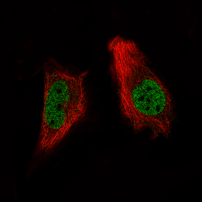
- Main image

- Experimental details
- Immunofluorescence staining of A-431 cells using the Anti-ARID1A monoclonal antibody, showing specific staining in the nucleoplasm in green. Microtubule- and nuclear probes are visualized in red and blue, respectively (where available).
- Sample type
- Human