Explore
 Validate
 Learn
Learn Western blot
Antibody data
- Antibody Data
- Antigen structure
- References [3]
- Comments [0]
- Validations
- Western blot [2]
- Immunohistochemistry [1]
- Flow cytometry [2]
Submit
Validation data
Reference
Comment
Report error
- Product number
- PA5-24186 - Provider product page

- Provider
- Invitrogen Antibodies
- Product name
- Folate Receptor alpha Polyclonal Antibody
- Antibody type
- Polyclonal
- Antigen
- Synthetic peptide
- Reactivity
- Human
- Host
- Rabbit
- Isotype
- IgG
- Vial size
- 400 μL
- Concentration
- 0.5 mg/mL
- Storage
- Store at 4°C short term. For long term storage, store at -20°C, avoiding freeze/thaw cycles.
Submitted references A patch of positively charged residues regulates the efficacy of clinical DR5 antibodies in solid tumors.
Unexpected PD-L1 immune evasion mechanism in TNBC, ovarian, and other solid tumors by DR5 agonist antibodies.
CD24-targeted intraoperative fluorescence image-guided surgery leads to improved cytoreduction of ovarian cancer in a preclinical orthotopic surgical model.
Shivange G, Mondal T, Lyerly E, Bhatnagar S, Landen CN, Reddy S, Kim J, Doan B, Riddle P, Tushir-Singh J
Cell reports 2021 Nov 2;37(5):109953
Cell reports 2021 Nov 2;37(5):109953
Unexpected PD-L1 immune evasion mechanism in TNBC, ovarian, and other solid tumors by DR5 agonist antibodies.
Mondal T, Shivange GN, Tihagam RG, Lyerly E, Battista M, Talwar D, Mosavian R, Urbanek K, Rashid NS, Harrell JC, Bos PD, Stelow EB, Stack MS, Bhatnagar S, Tushir-Singh J
EMBO molecular medicine 2021 Mar 5;13(3):e12716
EMBO molecular medicine 2021 Mar 5;13(3):e12716
CD24-targeted intraoperative fluorescence image-guided surgery leads to improved cytoreduction of ovarian cancer in a preclinical orthotopic surgical model.
Kleinmanns K, Fosse V, Davidson B, de Jalón EG, Tenstad O, Bjørge L, McCormack E
EBioMedicine 2020 Jun;56:102783
EBioMedicine 2020 Jun;56:102783
No comments: Submit comment
Supportive validation
- Submitted by
- Invitrogen Antibodies (provider)
- Main image
- Experimental details
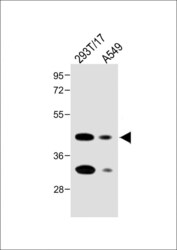
- Western blot analysis of Folate Receptor alpha in various lysates. Samples were incubated with Folate Receptor alpha polyclonal antibody (Product # PA5-24186) using a dilution of 1:2,000 followed by Goat Anti-Rabbit IgG, (H+L), Peroxidase conjugated at a dilution of 1:10,000. Lysates/proteins: 20 µg per lane. Lane 1: 293T/17 whole cell lysate; Lane 2: A549 whole cell lysate. Predicted band size: 30 kDa. Blocking/Dilution buffer: 5% NFDM/TBST.

- Submitted by
- Invitrogen Antibodies (provider)
- Main image
- Experimental details
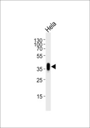
- Western blot analysis of Folate Receptor alpha in lysate from Hela cell line. Samples were incubated with Folate Receptor alpha polyclonal antibody (Product # PA5-24186) using a dilution of 1:1,000 followed by goat anti-rabbit IgG H&L (HRP) at a dilution of 1:5,000. Lysate at 35 µg.

Supportive validation
- Submitted by
- Invitrogen Antibodies (provider)
- Main image
- Experimental details
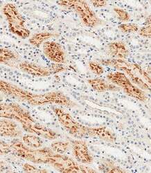
- Immunohistochemistry analysis of Folate Receptor alpha in paraffin-embedded Human kidney tissue. Samples were incubated with Folate Receptor alpha polyclonal antibody (Product # PA5-24186) using a dilution of 1:1,000 for 1 hours at room temperature followed by an undiluted biotinylated CRF Anti-Polyvalent HRP Polymer antibody.

Supportive validation
- Submitted by
- Invitrogen Antibodies (provider)
- Main image
- Experimental details
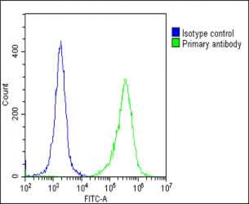
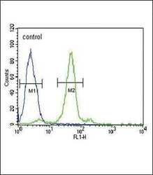
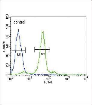
- Flow cytometry analysis of HeLa cells using a FOLR1 polyclonal antibody (Product # PA5-24186) (right) compared to a negative control cell (left) at a dilution of 1:10-50, followed by a FITC-conjugated goat anti-rabbit antibody
- Submitted by
- Invitrogen Antibodies (provider)
- Main image
- Experimental details
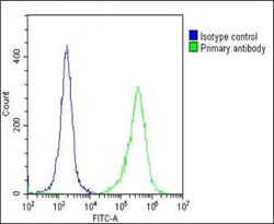
- Flow cytometry of (overlay histogram) of Folate Receptor alpha in Hela cells (green line). Samples were incubated with Folate Receptor alpha polyclonal antibody (Product # PA5-24186) using a dilution of 1:25 dilution for 60 min at 37°C followed by Goat-Anti-Rabbit IgG, DyLight® 488 Conjugated Highly Cross-Adsorbed at 1:200 dilution for 40 min at 37°C. The cells were fixed with 2% paraformaldehyde (10 min) and then permeabilized with 90% methanol for 10 min. The cells were then incubated in 2% bovine serum albumin to block non-specific protein-protein interactions followed by the primary antibody. Isotype control antibody (blue line) was rabbit IgG (1 μg/1x10^6 cells) used under the same conditions. Acquisition of >10, 000 events was performed.