 Explore
 Validate
 Learn
Learn Western blot
 ELISA
ELISAAntibody data
- Antibody Data
- Antigen structure
- References [4]
- Comments [0]
- Validations
- Western blot [1]
- Immunocytochemistry [1]
- Flow cytometry [2]
Submit
Validation data
Reference
Comment
Report error
- Product number
- MAB5646 - Provider product page

- Provider
- R&D Systems
- Product name
- Human FOLR1 Antibody
- Antibody type
- Monoclonal
- Description
- Protein A or G purified from hybridoma culture supernatant. Detects human FOLR1 in direct ELISAs and Western blots. In direct ELISAs, no cross-reactivity with recombinant human FOLR2, 3 or 4 is observed.
- Reactivity
- Human
- Host
- Mouse
- Conjugate
- Unconjugated
- Antigen sequence
P15328- Isotype
- IgG
- Antibody clone number
- 548908
- Vial size
- 100 ug
- Concentration
- LYOPH
- Storage
- Use a manual defrost freezer and avoid repeated freeze-thaw cycles. 12 months from date of receipt, -20 to -70 °C as supplied. 1 month, 2 to 8 °C under sterile conditions after reconstitution. 6 months, -20 to -70 °C under sterile conditions after reconstitution.
Submitted references Efficient delivery of small interfering RNAs targeting particular mRNAs into pancreatic cancer cells inhibits invasiveness and metastasis of pancreatic tumors.
Promising Nanocarriers for PEDF Gene Targeting Delivery to Cervical Cancer Cells Mediated by the Over-expressing FRα.
Conditions associated with circulating tumor-associated folate receptor 1 protein in healthy men and women.
Harnessing engineered antibodies of the IgE class to combat malignancy: initial assessment of FcɛRI-mediated basophil activation by a tumour-specific IgE antibody to evaluate the risk of type I hypersensitivity.
Taniuchi K, Yawata T, Tsuboi M, Ueba T, Saibara T
Oncotarget 2019 Apr 23;10(30):2869-2886
Oncotarget 2019 Apr 23;10(30):2869-2886
Promising Nanocarriers for PEDF Gene Targeting Delivery to Cervical Cancer Cells Mediated by the Over-expressing FRα.
Yang Y, He L, Liu Y, Xia S, Fang A, Xie Y, Gan L, He Z, Tan X, Jiang C, Tong A, Song X
Scientific reports 2016 Aug 31;6:32427
Scientific reports 2016 Aug 31;6:32427
Conditions associated with circulating tumor-associated folate receptor 1 protein in healthy men and women.
Kelemen LE, Brenton JD, Parkinson C, C Whitaker H, Piskorz AM, Csizmadi I, Robson PJ
PloS one 2014;9(5):e96542
PloS one 2014;9(5):e96542
Harnessing engineered antibodies of the IgE class to combat malignancy: initial assessment of FcɛRI-mediated basophil activation by a tumour-specific IgE antibody to evaluate the risk of type I hypersensitivity.
Rudman SM, Josephs DH, Cambrook H, Karagiannis P, Gilbert AE, Dodev T, Hunt J, Koers A, Montes A, Taams L, Canevari S, Figini M, Blower PJ, Beavil AJ, Nicodemus CF, Corrigan C, Kaye SB, Nestle FO, Gould HJ, Spicer JF, Karagiannis SN
Clinical and experimental allergy : journal of the British Society for Allergy and Clinical Immunology 2011 Oct;41(10):1400-13
Clinical and experimental allergy : journal of the British Society for Allergy and Clinical Immunology 2011 Oct;41(10):1400-13
No comments: Submit comment
Supportive validation
- Submitted by
- R&D Systems (provider)
- Main image
- Experimental details
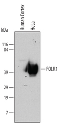
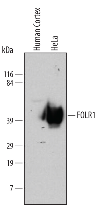
- Detection of Human FOLR1 by Western Blot. Western blot shows lysates of human cortex tissue and HeLa human cervical epithelial carcinoma cell line. PVDF membrane was probed with 2 µg/mL of Mouse Anti-Human FOLR1 Monoclonal Antibody (Catalog # MAB5646) followed by HRP-conjugated Anti-Mouse IgG Secondary Antibody (Catalog # HAF007). A specific band was detected for FOLR1 at approximately 40 kDa (as indicated). This experiment was conducted using Immunoblot Buffer Group 1. Use under non-reducing conditions only.
Supportive validation
- Submitted by
- R&D Systems (provider)
- Main image
- Experimental details
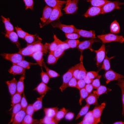
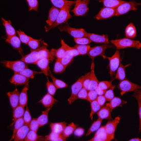
- FOLR1 in MCF-7 Human Cell Line. FOLR1 was detected in immersion fixed MCF-7 human breast cancer cell line using 10 µg/mL Mouse Anti-Human FOLR1 Monoclonal Antibody (Catalog # MAB5646) for 3 hours at room temperature. Cells were stained with the NorthernLights™ 557-conjugated Anti-Mouse IgG Secondary Antibody (red; Catalog # NL007) and counterstained with DAPI (blue). View our protocol for Fluorescent ICC Staining of Cells on Coverslips.
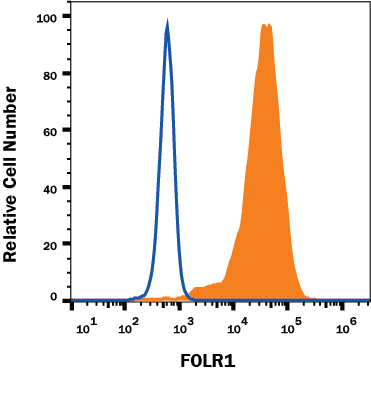
Supportive validation
- Submitted by
- R&D Systems (provider)
- Main image

- Experimental details
- Detection of FOLR1 in MCF-7 Human Cell Line by Flow Cytometry. MCF-7 human breast cancer cell line was stained with Mouse Anti-Human FOLR1 Monoclonal Antibody (Catalog # MAB5646, filled histogram) or isotype control antibody (Catalog # MAB002, open histogram), followed by Phycoerythrin-conjugated Anti-Mouse IgG Secondary Antibody (Catalog # F0102B). View our protocol for Staining Membrane-associated Proteins.
- Submitted by
- R&D Systems (provider)
- Main image
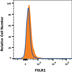
- Experimental details
- FOLR1 Specificity is Shown by Flow Cytometry in Knockout Cell Line. FOLR1 knockout MCF-7 human breast cancer cell line was stained with Mouse Anti-Human FOLR1 Monoclonal Antibody (Catalog # MAB5646, filled histogram) or isotype control antibody (Catalog # MAB002, open histogram) followed by anti-Mouse IgG PE-conjugated secondary antibody (Catalog # F0102B). No staining in the FOLR1 knockout MCF-7 cell line was observed. View our protocol for Staining Membrane-associated Proteins.
