 Explore
 Validate
 Learn
Learn Western blot
Antibody data
- Antibody Data
- Antigen structure
- References [0]
- Comments [0]
- Validations
- Western blot [2]
- Immunocytochemistry [2]
- Immunohistochemistry [4]
Submit
Validation data
Reference
Comment
Report error
- Product number
- PA5-52147 - Provider product page

- Provider
- Invitrogen Antibodies
- Product name
- SEC31A Polyclonal Antibody
- Antibody type
- Polyclonal
- Antigen
- Recombinant full-length protein
- Description
- Immunogen sequence: NWREALAAVL TYAKPDEFSA LCDLLGTRLE NEGDSLLQTQ ACLCYICAGN VEKLVACWTK AQDGSHPLSL QDLIEKVVIL RKAVQLTQAM DTSTVGVLLA AKMSQYANLL AAQGSIAAAL AFLPDNTNQP NIMQLR Highest antigen sequence identity to the following orthologs: Mouse - 93%, Rat - 92%.
- Reactivity
- Human
- Host
- Rabbit
- Isotype
- IgG
- Vial size
- 100 µL
- Concentration
- 0.2 mg/mL
- Storage
- Store at 4°C short term. For long term storage, store at -20°C, avoiding freeze/thaw cycles.
No comments: Submit comment
Supportive validation
- Submitted by
- Invitrogen Antibodies (provider)
- Main image

- Experimental details
- Knockdown of SEC31A was achieved by transfecting Hep G2 with SEC31A specific siRNAs (Silencer® select Product # s22550, s22551). Western blot analysis (Fig. a) was performed using membrane enriched cell extracts from the SEC31A knockdown cells (Lane 3), non-specific scrambled siRNA transfected cells (Lane 2) and untransfected cells (Lane 1). The blot was probed with SEC31A Polyclonal Antibody (Product # PA5-52147, 0.4 µg/ml) and Goat anti-Rabbit IgG (H+L) Superclonal™ Secondary Antibody, HRP conjugate (Product # A27036, 0.25µg/ml, 1:4000 dilution). Densitometric analysis of this western blot is shown in histogram (Fig. b). Decrease in signal upon siRNA mediated knock down confirms that antibody is specific to SEC31A.

- Submitted by
- Invitrogen Antibodies (provider)
- Main image

- Experimental details
- Western blot was performed using Anti-SEC31A Rabbit Polyclonal Antibody (Product # PA5-52147) and a 110 kDa band corresponding to SEC31A was observed across cell lines tested. Membrane enriched cell extracts (30 µg lysate) of A-431 (Lane 1), A549 (Lane 2), Hep G2 (Lane 3), K-562 (Lane 4), and SH-SY5Y (Lane 5) were electrophoresed using Novex® NuPAGE® 4-12 % Bis-Tris gel (Product # NP0321BOX). Resolved proteins were then transferred onto a nitrocellulose membrane (Product # IB23001) by iBlot® 2 Dry Blotting System (Product # IB21001). The blots were probed with the primary antibody (0.4 µg/ml), Goat Anti-Rabbit IgG Secondary Antibody, HRP conjugate (Product # A27036, 1:4000 dilution) using the iBright FL 1000 (Product # A32752). Chemiluminescent detection was performed using SuperSignal™ West Dura Extended Duration Substrate (Product # 34076).

Supportive validation
- Submitted by
- Invitrogen Antibodies (provider)
- Main image

- Experimental details
- Immunofluorescent staining of SEC31A in human cell line U-251 MG shows positivity in cytoplasm & vesicles. Samples were probed using a SEC31A Polyclonal Antibody (Product # PA5-52147).

- Submitted by
- Invitrogen Antibodies (provider)
- Main image

- Experimental details
- Immunofluorescence analysis of SEC31A was performed using 70% confluent log phase A-431 cells. The cells were fixed with 4% paraformaldehyde for 10 minutes, permeabilized with 0.1% Triton™ X-100 for 15 minutes, and blocked with 2% BSA for 1 hour at room temperature. The cells were labeled with SEC31A Polyclonal Antibody (Product # PA5-52147) at 2 µg/mL in 0.1% BSA, incubated at 4 degree Celsius overnight and then with Goat anti-Rabbit IgG (H+L) Superclonal™ Secondary Antibody, Alexa Fluor® 488 conjugate (Product # A27034) at a dilution of 1:2000 for 45 minutes at room temperature (Panel a: green). Nuclei (Panel b: blue) were stained with SlowFade® Gold Antifade Mountant with DAPI (Product # S36938). F-actin (Panel c: red) was stained with Rhodamine Phalloidin (Product # R415, 1:300). Panel d represents the merged image showing staining in membrane and cytoplasm. Panel e represents control cells with no primary antibody to assess background. The images were captured at 60X magnification.

Supportive validation
- Submitted by
- Invitrogen Antibodies (provider)
- Main image
- Experimental details
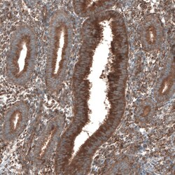
- Immunohistochemical staining of SEC31A in human endometrium using SEC31A Polyclonal Antibody (Product # PA5-52147) shows positivit in glandular cells.

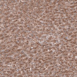
- Submitted by
- Invitrogen Antibodies (provider)
- Main image
- Experimental details
- Immunohistochemical staining of SEC31A in human cerebral cortex using SEC31A Polyclonal Antibody (Product # PA5-52147) shows positivity in neurons.

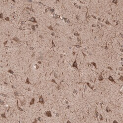
- Submitted by
- Invitrogen Antibodies (provider)
- Main image
- Experimental details
- Immunohistochemical staining of SEC31A in human liver using SEC31A Polyclonal Antibody (Product # PA5-52147) shows positivity in hepatocytes.

- Submitted by
- Invitrogen Antibodies (provider)
- Main image
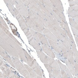
- Experimental details
- Immunohistochemical staining of SEC31A in human skeletal muscle using SEC31A Polyclonal Antibody (Product # PA5-52147) shows very weak positivity in myocytes.
