 Explore
 Validate
 Learn
Learn Western blot
 Gel shift
Antibody data
- Antibody Data
- Antigen structure
- References [4]
- Comments [0]
- Validations
- Western blot [2]
- Immunocytochemistry [2]
- Other assay [2]
Submit
Validation data
Reference
Comment
Report error
- Product number
- PA3-023 - Provider product page

- Provider
- Invitrogen Antibodies
- Product name
- Cyclophilin 40 Polyclonal Antibody
- Antibody type
- Polyclonal
- Antigen
- Recombinant full-length protein
- Description
- PA3-023 detects cyclophilin 40 (CyP-40) from human, mouse and rat tissues and cells. PA3-023 has been successfully used in Western blot and gel shift procedures. By Western blot, this antibody detects a 40 kDa protein representing CyP-40 from rat brain extract. The PA3-023 immunogen is recombinant human CyP-40 expressed in E. coli.
- Reactivity
- Human, Mouse, Rat
- Host
- Rabbit
- Isotype
- IgG
- Vial size
- 100 µL
- Concentration
- Conc. Not Determined
- Storage
- -20° C, Avoid Freeze/Thaw Cycles
Submitted references A comparison of Hsp90alpha and Hsp90beta interactions with cochaperones and substrates.
Cyclophilin A is required for retinoic acid-induced neuronal differentiation in p19 cells.
Multiple components of the HSP90 chaperone complex function in regulation of heat shock factor 1 In vivo.
Multiple components of the HSP90 chaperone complex function in regulation of heat shock factor 1 In vivo.
Taherian A, Krone PH, Ovsenek N
Biochemistry and cell biology = Biochimie et biologie cellulaire 2008 Feb;86(1):37-45
Biochemistry and cell biology = Biochimie et biologie cellulaire 2008 Feb;86(1):37-45
Cyclophilin A is required for retinoic acid-induced neuronal differentiation in p19 cells.
Song J, Lu YC, Yokoyama K, Rossi J, Chiu R
The Journal of biological chemistry 2004 Jun 4;279(23):24414-9
The Journal of biological chemistry 2004 Jun 4;279(23):24414-9
Multiple components of the HSP90 chaperone complex function in regulation of heat shock factor 1 In vivo.
Bharadwaj S, Ali A, Ovsenek N
Molecular and cellular biology 1999 Dec;19(12):8033-41
Molecular and cellular biology 1999 Dec;19(12):8033-41
Multiple components of the HSP90 chaperone complex function in regulation of heat shock factor 1 In vivo.
Bharadwaj S, Ali A, Ovsenek N
Molecular and cellular biology 1999 Dec;19(12):8033-41
Molecular and cellular biology 1999 Dec;19(12):8033-41
No comments: Submit comment
Supportive validation
- Submitted by
- Invitrogen Antibodies (provider)
- Main image

- Experimental details
- Knockdown of Cyclophilin 40 was achieved by transfecting LNCaP with Cyclophilin 40 specific siRNAs (Silencer® select Product # S10913, S10915). Western blot analysis (Fig. a) was performed using Whole cell extracts from the Cyclophilin 40 knockdown cells (lane 3), non-targeting scrambled siRNA transfected cells (lane 2) and untransfected cells (lane 1). The blot was probed with Cyclophilin 40 Polyclonal Antibody (Product # PA3-023, 1:1000 dilution) and Goat anti-Rabbit IgG (H+L) Superclonal™ Recombinant Secondary Antibody, HRP (Product # A27036, 1:20,000 dilution). Densitometric analysis of this western blot is shown in histogram (Fig. b). Decrease in signal upon siRNA mediated knock down confirms that antibody is specific to Cyclophilin 40.

- Submitted by
- Invitrogen Antibodies (provider)
- Main image
- Experimental details
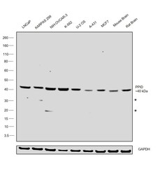
- Western blot was performed using Anti-Cyclophilin 40 Polyclonal Antibody (Product # PA3-023) and a ~40 kDa band corresponding to Cyclophilin 40 was observed across cell lines and tissues tested. Whole cell lysate (40 µg lysate) of LNCaP (Lane 1), KARPAS 299 (Lane 2), NIH:OVCAR-3 (Lane 3), K-562 (Lane 4), U-2 OS (Lane 5), A-431 (Lane 6), MCF7 (Lane 7), Mouse Brain (Lane 8) and Rat Brain (Lane 9) were electrophoresed using NuPAGE™ 4-12% Bis-Tris Protein Gel (Product # NP0321BOX), 10 well. Resolved proteins were then transferred onto a nitrocellulose membrane (Product # IB23001) by iBlot® 2 Dry Blotting System (Product # IB21001). The blot was probed with the primary antibody (1:1000 dilution) and detected by chemiluminescence with Goat anti-Rabbit IgG (H+L) Superclonal™ Recombinant Secondary Antibody, HRP (Product # A27036, 1:20,000 dilution) using the iBright™ FL1500 Imaging System (Product # A44115). Chemiluminescent detection was performed using SuperSignal™ West Dura Extended Duration Substrate (Product # 34076).

Supportive validation
- Submitted by
- Invitrogen Antibodies (provider)
- Main image

- Experimental details
- Immunofluorescence analysis of Cyclophilin D was done on 70% confluent log phase A431 cells. The cells were fixed with 4% paraformaldehyde for 10 minutes, permeabilized with 0.1% Triton™ X-100 for 10 minutes, and blocked with 1% BSA for 1 hour at room temperature. The cells were labeled with Cyclophilin D Rabbit Polyclonal Antibody (Product # PA3-023) at 1:250 dilution in 0.1% BSA and incubated for 3 hours at room temperature and then labeled with Goat anti-Rabbit IgG (H+L) Superclonal™ Secondary Antibody, Alexa Fluor® 488 conjugate (Product # A27034) at a dilution of 1:2000 for 45 minutes at room temperature (Panel a: green). Nuclei (Panel b: blue) were stained with SlowFade® Gold Antifade Mountant with DAPI (Product # S36938). F-actin (Panel c: red) was stained with Rhodamine Phalloidin (Product # R415, 1:300). Panel d is a merged image showing cytoplasmic localization. Panel e is a no primary antibody control. The images were captured at 60X magnification.

- Submitted by
- Invitrogen Antibodies (provider)
- Main image

- Experimental details
- Immunofluorescence analysis of Cyclophilin 40 was performed using 70% confluent log phase A549 cells. The cells were fixed with 4% paraformaldehyde for 10 minutes, permeabilized with 0.01% Triton™ X-100 for 10 minutes, and blocked with 2% BSA for 1 hour at room temperature. The cells were labeled with Cyclophilin 40 Polyclonal Antibody (Product # PA3-023) at 1:100 dilution in 0.1% BSA, incubated at 4 degree celsius overnight and then labeled with Goat anti-Rabbit IgG (H+L) Superclonal™ Recombinant Secondary Antibody, Alexa Fluor® 488 conjugate (Product # A27034, 1:2000 dilution), for 45 minutes at room temperature (Panel a: Green). Nuclei (Panel b:Blue) were stained with Hoechst 33342 (Product # H1399). F-actin (Panel c: Red) was stained with Alexa Fluor™ Plus 647 Phalloidin (Product # A30107, 1:2000 dilution). Panel d represents the merged image showing Nucleus and cytoplasm localization. Panel e represents control cells with no primary antibody to assess background. The images were captured at 40x magnification in CellInsight CX7 LZR High-Content Screening (HCS) Platform (Product # CX7A1110LZR) and externally deconvoluted (D.Sage et al./Methods 115 (2017) 28–41.

Supportive validation
- Submitted by
- Invitrogen Antibodies (provider)
- Main image

- Experimental details
- NULL

- Submitted by
- Invitrogen Antibodies (provider)
- Main image

- Experimental details
- NULL
