 Explore
 Validate
 Learn
Learn Western blot
 ELISA
ELISAAntibody data
- Antibody Data
- Antigen structure
- References [0]
- Comments [0]
- Validations
- Western blot [3]
- Immunocytochemistry [1]
- Immunohistochemistry [2]
Submit
Validation data
Reference
Comment
Report error
- Product number
- F40119 - Provider product page

- Provider
- NSJ Bioreagents
- Product name
- SYVN1 Antibody (HRD1)
- Antibody type
- Polyclonal
- Description
- This highly specific SYVN1 antibody is suitable for use in Immunofluorescence/Immunohistochemistry/Western blot/ELISA applications with human and mouse samples.
- Reactivity
- Human, Mouse
- Host
- Rabbit
- Conjugate
- Unconjugated
- Vial size
- 0.08 ml, 0.4 ml
- Concentration
- In 1X PBS, pH 7.4, with 0.09% sodium azide
- Storage
- Aliquot the SYVN1 antibody and store frozen at -20oC or colder. Avoid repeated freeze-thaw cycles.
No comments: Submit comment
Supportive validation
- Submitted by
- NSJ Bioreagents (provider)
- Main image
- Experimental details
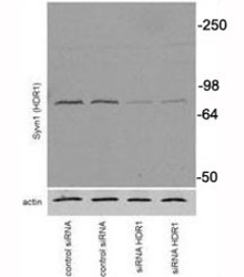
- Western blot testing of SYVN1 antibody and transiently transfected Neuro2A cells

- Submitted by
- NSJ Bioreagents (provider)
- Main image

- Experimental details
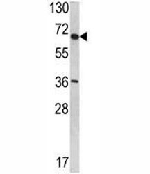
- Western blot analysis of SYVN1 antibody in T47D lysate
- Submitted by
- NSJ Bioreagents (provider)
- Main image
- Experimental details
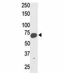
- Western blot analysis of SYVN1 mouse kidney tissue lysate

Supportive validation
- Submitted by
- NSJ Bioreagents (provider)
- Main image

- Experimental details
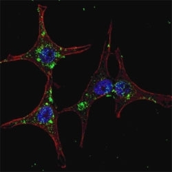
- Fluorescent confocal image of HeLa cells stained with SYVN1 antibody. Primary Ab (1:200, 2 h at room temperature); Alexa Fluor 488 conjugated donkey anti-rabbit Ab (green) was used as secondary (1:1000, 1h). Nuclei were counterstained with Hoechst 33342 (blue) (10 ug/ml, 5 min).
Supportive validation
- Submitted by
- NSJ Bioreagents (provider)
- Main image
- Experimental details
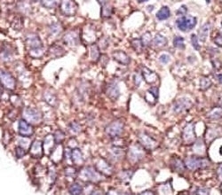
- IHC analysis of FFPE human breast carcinoma tissue stained with the SYVN1 antibody

- Submitted by
- NSJ Bioreagents (provider)
- Main image
- Experimental details
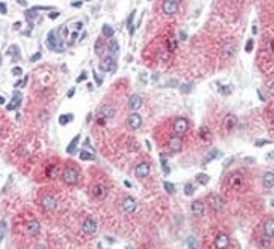
- IHC analysis of FFPE human liver tissue stained with SYVN1 antibody
