Explore
 Validate
 Learn
Learn Western blot
 Immunocytochemistry
Antibody data
- Antibody Data
- Antigen structure
- References [0]
- Comments [0]
- Validations
- Western blot [3]
- Immunohistochemistry [3]
Submit
Validation data
Reference
Comment
Report error
- Product number
- LS-C99633 - Provider product page

- Provider
- LSBio
- Product name
- SYVN1 / HRD1 Antibody (aa586-617) LS-C99633
- Antibody type
- Polyclonal
- Description
- Ammonium sulfate precipitation
- Reactivity
- Human, Mouse
- Host
- Rabbit
- Storage
- Maintain refrigerated at 2°C to 8°C for up to 6 months. For long term storage store at -20°C.
No comments: Submit comment
Supportive validation
- Submitted by
- LSBio (provider)
- Main image
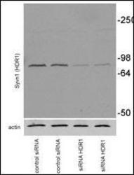
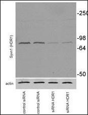
- Experimental details
- Mouse Neuroblastoma Neuro2A (N2A) was transiently transfected, collected at 72h after transfection. Primary antibodies against syvn1 (1:1000) and anti-rabbit secondary POD-conjugated antibodies from Pierce Biotechnology, Inc (Rockford, IL, 1:2000)(Provided by Dr. Susana Granell & Institution University of Arkansas).
- Submitted by
- LSBio (provider)
- Main image

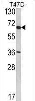
- Experimental details
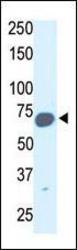
- Western blot of SYVN1 (HRD1) Antibody in T47D cell line lysates (35 ug/lane). HRD1 (arrow) was detected using the purified antibody.
- Submitted by
- LSBio (provider)
- Main image
- Experimental details
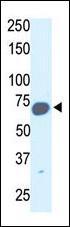
- The anti-HRD1 antibody is used in Western blot to detect HRD1 in mouse kidney tissue lysate.
Supportive validation
- Submitted by
- LSBio (provider)
- Main image
- Experimental details
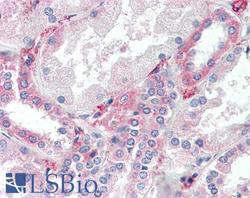
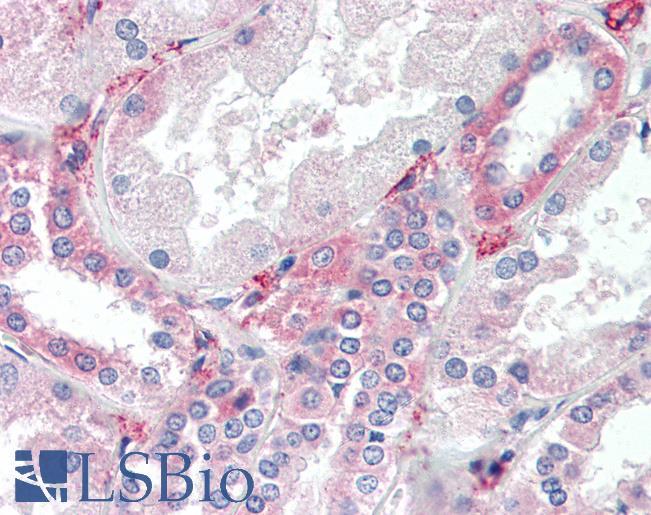
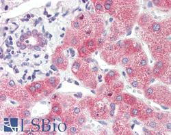
- Anti-SYVN1 / HRD1 antibody IHC of human kidney. Immunohistochemistry of formalin-fixed, paraffin-embedded tissue after heat-induced antigen retrieval. Antibody concentration 10 ug/ml.
- Submitted by
- LSBio (provider)
- Main image
- Experimental details
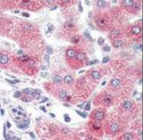
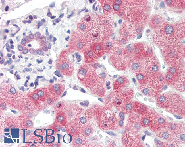
- Anti-SYVN1 / HRD1 antibody IHC of human liver. Immunohistochemistry of formalin-fixed, paraffin-embedded tissue after heat-induced antigen retrieval. Antibody concentration 10 ug/ml.
- Submitted by
- LSBio (provider)
- Main image

- Experimental details
- Formalin-fixed and paraffin-embedded human Liver tissue reacted with SYVN1 (HRD1) Antibody , which was peroxidase-conjugated to the secondary antibody, followed by AEC staining. This data demonstrates the use of this antibody for immunohistochemistry; clinical relevance has not been evaluated.