Explore
 Validate
 Learn
Learn Western blot
 Immunocytochemistry
Antibody data
- Antibody Data
- Antigen structure
- References [0]
- Comments [0]
- Validations
- Immunocytochemistry [2]
- Immunohistochemistry [1]
Submit
Validation data
Reference
Comment
Report error
- Product number
- PA1-46121 - Provider product page

- Provider
- Invitrogen Antibodies
- Product name
- HRD1 Polyclonal Antibody
- Antibody type
- Polyclonal
- Antigen
- Synthetic peptide
- Description
- Suggested positive control: 293T, INS-1, and MIN6 cell lysates, antigen standard for SYVN1 (transient overexpression lysate).
- Reactivity
- Human, Mouse, Hamster
- Host
- Rabbit
- Isotype
- IgG
- Vial size
- 100 μL
- Concentration
- 1.0 mg/mL
- Storage
- -20°C, Avoid Freeze/Thaw Cycles
No comments: Submit comment
Supportive validation
- Submitted by
- Invitrogen Antibodies (provider)
- Main image

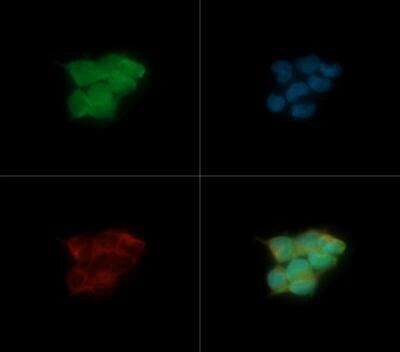
- Experimental details
- Immunocytochemistry analysis of HRD1 in HEK-293 cells. Samples were incubated in HRD1 polyclonal antibody (Product # PA1-46121) followed by DyLight 488 (green). Nuclei and alpha-tubulin were counterstained with DAPI (blue) and DyLight 550 (red).
- Submitted by
- Invitrogen Antibodies (provider)
- Main image

- Experimental details
- Immunocytochemistry analysis of HRD1 in HEK-293 cells. Samples were incubated in HRD1 polyclonal antibody (Product # PA1-46121) followed by DyLight 488 (green). Nuclei and alpha-tubulin were counterstained with DAPI (blue) and DyLight 550 (red).

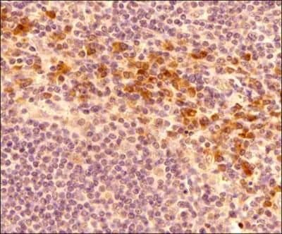
Supportive validation
- Submitted by
- Invitrogen Antibodies (provider)
- Main image

- Experimental details
- Immunohistochemical analysis of HRD1 in formalin-fixed paraffin-embedded tissue section of human tonsil. Samples were incubated in HRD1 polyclonal antibody (Product # PA1-46121) using a dilution of 1:500 followed by a HRP conjugated anti-Rabbit secondary antibody with DAB reagent. The sections were further counterstained with hematoxylin for labeling cellular nuclei. This HRD1 antibody generated an expected cytoplasmic staining of HRD1 with stronger signal around periphery of the nucleus signifying the staining of the endoplasmic reticulum.