Explore
 Validate
 Learn
Learn Western blot
Antibody data
- Antibody Data
- Antigen structure
- References [0]
- Comments [0]
- Validations
- Western blot [1]
- Immunocytochemistry [1]
- Immunohistochemistry [1]
Submit
Validation data
Reference
Comment
Report error
- Product number
- ANR-021-200UL - Provider product page

- Provider
- Invitrogen Antibodies
- Product name
- NPY1R Polyclonal Antibody
- Antibody type
- Polyclonal
- Antigen
- Other
- Reactivity
- Human, Rat
- Host
- Rabbit
- Isotype
- IgG
- Vial size
- 200 µL
- Concentration
- 0.75 mg/mL
- Storage
- -20° C, Avoid Freeze/Thaw Cycles
No comments: Submit comment
Supportive validation
- Submitted by
- Invitrogen Antibodies (provider)
- Main image

- Experimental details
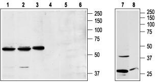
- Western blot analysis of human Jurkat acute T cell leukemia (lanes 1 and 4), human K562 chronic myelogeneous leukemia (lanes 2 and 5), and rat RBL basophilic leukemia (lanes 3 and 6) cell lysates and rat brain lysates (lanes 7 and 8): - 1,2,3,7. Anti-NPY1R Antibody (#ANR-021), (1:200).4,5,6,8. Anti-NPY1R Antibody , preincubated with NPY1R Blocking Peptide (#BLP-NR021).
Supportive validation
- Submitted by
- Invitrogen Antibodies (provider)
- Main image

- Experimental details
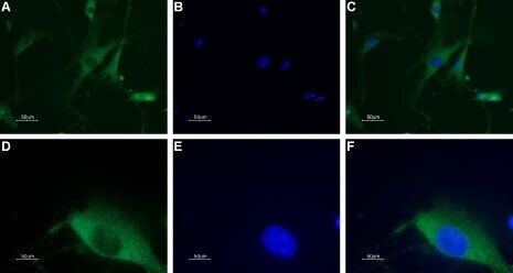
- Expression of neuropeptide Y1 receptor in DRG neurons - Immunocytochemical staining of a primary culture of rat dorsal root ganglion (DRG) neurons. A, D. A paraformaldehyde-fixed and permeabilized DRG primary culture was stained with Anti-NPY1R Antibody (#ANR-021), (1:100), followed by Alexa-555-conjugated goat- Anti-rabbit secondary Antibody . B, E. Nuclear fluorescence staining of cells using the membrane-permeable DNA dye Hoechst 33342. C. Merged images of A and B. F.Merged images of D and E. Magnification:A-C: x20E-F: x100.
Supportive validation
- Submitted by
- Invitrogen Antibodies (provider)
- Main image
- Experimental details
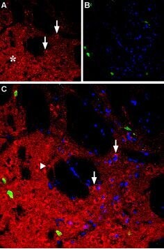
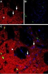
- Expression of neuropeptide Y1 receptor in rat striatum - Immunohistochemical staining of rat striatum using Anti-NPY1R Antibody (#ANR-021). A. NPY1R (red) appears in the striatal matrix (white asterisk) and in medium-size cells in the matrix (arrows). B. Parvalbumin (green) appears in the striatal matrix. C. Confocal merge of NPY1R and parvalbumin indicates that NPY1R is restricted to granule cells. DAPI is used as the counterstain (blue).