Explore
 Validate
 Learn
LearnPA5-42058
antibody from Invitrogen Antibodies
Targeting: HSD17B1
EDH17B2, EDHB17, HSD17, MGC138140, SDR28C1
 Western blot
 Immunohistochemistry
Antibody data
- Antibody Data
- Antigen structure
- References [2]
- Comments [0]
- Validations
- Immunohistochemistry [1]
- Other assay [5]
Submit
Validation data
Reference
Comment
Report error
- Product number
- PA5-42058 - Provider product page

- Provider
- Invitrogen Antibodies
- Product name
- HSD17B1 Polyclonal Antibody
- Antibody type
- Polyclonal
- Antigen
- Synthetic peptide
- Description
- Peptide sequence: MARTVVLITG CSSGIGLHLA VRLASDPSQS FKVYATLRDL KTQGRLWEAA Sequence homology: Cow: 100%; Dog: 100%; Guinea Pig: 91%; Horse: 100%; Human: 100%; Mouse: 100%; Pig: 100%; Rabbit: 100%; Rat: 100%
- Reactivity
- Human, Mouse
- Host
- Rabbit
- Isotype
- IgG
- Vial size
- 100 μL
- Concentration
- 0.5 mg/mL
- Storage
- -20°C, Avoid Freeze/Thaw Cycles
Submitted references Platelets induce increased estrogen production through NF-κB and TGF-β1 signaling pathways in endometriotic stromal cells.
Estrogen biosynthesis in cultured skeletal muscle cells (L6) induced by amino acids.
Qi Q, Liu X, Zhang Q, Guo SW
Scientific reports 2020 Jan 28;10(1):1281
Scientific reports 2020 Jan 28;10(1):1281
Estrogen biosynthesis in cultured skeletal muscle cells (L6) induced by amino acids.
Iresjö BM, Landin A, Ohlsson C, Lundholm K
Genes & nutrition 2019;14:29
Genes & nutrition 2019;14:29
No comments: Submit comment
Supportive validation
- Submitted by
- Invitrogen Antibodies (provider)
- Main image

- Experimental details
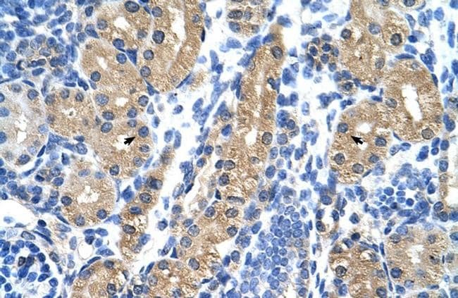
- Immunohistochemistry (paraffin-embedded) analysis of human kidney tissue using an anti-HSD17B1 polyclonal antibody (Product # PA5-42058).
Supportive validation
- Submitted by
- Invitrogen Antibodies (provider)
- Main image
- Experimental details
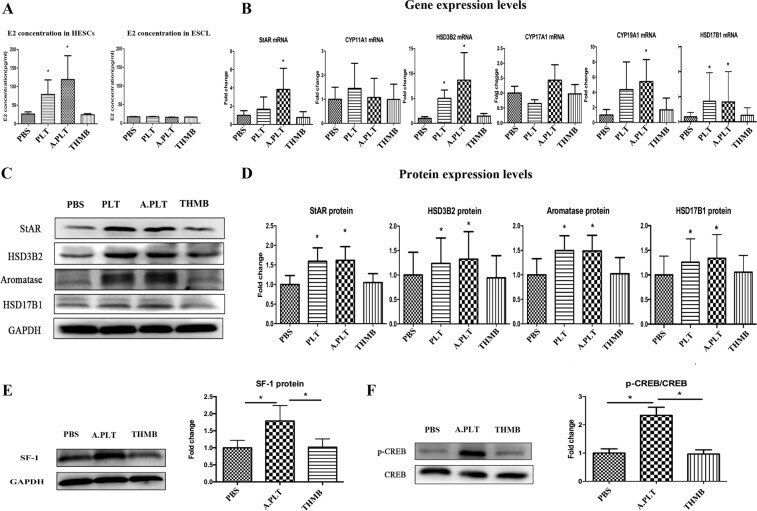
- Figure 1 Treatment of HESCs with activated platelets resulted in increased production of E 2 through upregulation of steroidogenic genes. ( A ) Concentrations of E 2 in the supernatant of HESCs (n = 7) and ESCL (n = 5) treated with PBS, platelets, activated platelets, and thrombin alone. Gene ( B ) and protein ( D ) expression levels of the genes involved in estrogen biosynthesis: StAR, CYP11A1, HSD3B2, CYP17A1, CYP19A1 and HSD17B1 in HESCs co-cultured with PBS, platelets, activated platelets and thrombin alone. ( C ) Representative Western blotting results for StAR, HSD3B2, aromatase and HSD17B1. Fold change for SF-1 ( E ) and p-CREB ( F ) protein expression in HESCs co-cultured with PBS, activated platelets and thrombin alone. ""*"" denotes that the p-value of the difference between the designated treatment and the PBS treatment is less than 0.05. Abbreviations used: PLT: platelets; A.PLT: activated platelets; THMB: thrombin. Paired Wilcoxon's test was used.
- Submitted by
- Invitrogen Antibodies (provider)
- Main image

- Experimental details
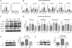
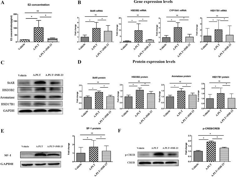
- Figure 3 Effect of suppression of NF-kappaB activation by JSH-23 on the production of E 2 and expression of steroidogenic genes in HESCs. ( A ) Concentrations of E 2 in the supernatant of HESCs (n = 8) treated with vehicle, activated platelets and activated platelets + JSH-23. ( B ) Gene expression levels of the genes involved in estrogen biosynthesis StAR, HSD3B2, CYP19A1 and HSD17B1 in HESCs co-cultured with vehicle, activated platelets and activated platelets + JSH-23. ( C ) Representative Western blotting results for StAR, HSD3B2, aromatase and HSD17B1 protein expression. ( D ) Summary of fold changes of protein expression levels of StAR, HSD3B2, aromatase and HSD17B1 in HESCs (n = 8) treated with vehicle, activated platelets and activated platelets + JSH-23. Summary of fold changes for SF-1 ( E ) and p-CREB ( F ) protein expression in HESCs (n = 8) treated with vehicle, activated platelets and activated platelets + JSH-23. ""*"" denotes that the p-value of the difference between the groups is less than 0.05. NS: p > 0.05. Paired Wilcoxon's test was used.
- Submitted by
- Invitrogen Antibodies (provider)
- Main image

- Experimental details
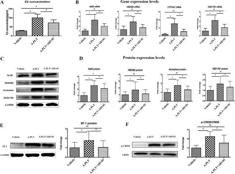
- Figure 5 Effects of neutralization of TGF-beta1 by A83-01 on the production of E 2 and expression of genes involved in steroidogenesis in HESCs. ( A ) Concentrations of E 2 in the supernatant of HESCs (n = 8) treated with vehicle, activated platelets and activated platelets + A83-01. ( B ) Gene expression levels of StAR, HSD3B2, CYP19A1 and HSD17B1 in HESCs (n = 8) treated with vehicle, activated platelets and activated platelets + A83-01. ( C ) Representative Western blotting results for StAR, HSD3B2, aromatase and HSD17B1 in HESCs under different treatments. ( D ) Summary of fold changes of protein expression levels of StAR, HSD3B2, aromatase and HSD17B1 in HESCs (n = 8) treated with vehicle, activated platelets and activated platelets + A83-01. Western blot analysis results for SF-1 ( E ) and p-CREB ( F ) proteins in HESCs treated with vehicle, activated platelets and activated platelets + A83-01. ""*"" denotes that the p-value of the difference between the groups is less than 0.05. NS: p > 0.05. Paired Wilcoxon's test was used.
- Submitted by
- Invitrogen Antibodies (provider)
- Main image
- Experimental details
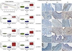
- Figure 6 Effect of non-immune serum (NI), platelet infusion (PI) and platelet depletion (PD) on lesional development and lesional expression of steroidogenic proteins in mice with induced endometriosis. ( A ) Change in hotplate latency for mice in the NI, PD and PI groups tested at the time indicated. ( B ) Total lesion weight for mice in the 3 treatment groups. Boxplots of lesional staining levels in NI, PD and PI groups: StAR ( C ), HSD3B2 ( D ), aromatase ( E ), HSD17B1 ( F ), SF-1 ( G ), and p-CREB ( H ). The dotted horizontal line represents the median of all 3 groups. Symbols of statistical significance: *: p < 0.05, **:p < 0.01, ***: p < 0.001. n = 10 for each group. Except in ( A ), where the comparison was made using Kruskal's test, all tests were made using Wilcoxon's test in comparison with the NI group. ( I ) Representative photomicrographs of StAR, HSD3B2, aromatase, HSD17B1, SF-1 and p-CREB staining in endometriotic lesions (x400) in Balb/c mice with induced endometriosis in different treatment groups. Scale bar = 50 mum.

- Submitted by
- Invitrogen Antibodies (provider)
- Main image
- Experimental details
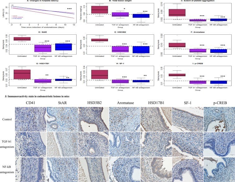
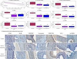
- Figure 7 Effect of antagonism of TGF-beta1 (by SB431542) or of NF-kappaB (by JSH-23) on lesional development and lesional expression of steroidogenic proteins in mice with induced endometriosis. ( A ) Change in hotplate latency for mice treatment with vehicle (untreated, or U), antagonism of TGF-beta1 (T), and antagonism of NF-kappaB (N) tested at the time indicated. ( B ) Total lesion weight in the 3 groups of mice. ( C ) Boxplots of the extent of lesional platelet aggregation using CD41 the 3 groups of mice. Boxplots of lesional staining levels in the 3 groups of mice: StAR ( D ), HSD3B2 ( E ), aromatase ( F ), HSD17B1 ( G ), SF-1 ( H ), and p-CREB ( I ). The dotted horizontal line represents the median of all 3 groups. Symbols of statistical significance: *: p < 0.05, **: p < 0.01, ***: p < 0.001. NS: p > 0.05. n = 10 for each group. Except in ( A ), where the comparison was made using Kruskal's test, all tests were made using Wilcoxon's test in comparison with the untreated group ( U ). ( J ) Representative immunoreactivity staining of CD41, StAR, HSD3B2, aromatase, HSD17B1, SF-1 and p-CREB in the endometriotic lesions (x400) in Balb/c mice with induced endometriosis which then treatment with vehicle, SB431542 (an antagonist for TGF-beta1) and JSH-23 (an antagonist for NF-kappaB) for two weeks. Scale bar = 50 mum.