Explore
 Validate
 Learn
Learn Western blot
 Immunohistochemistry
Antibody data
- Antibody Data
- Antigen structure
- References [2]
- Comments [0]
- Validations
- Immunohistochemistry [20]
- Other assay [2]
Submit
Validation data
Reference
Comment
Report error
- Product number
- MA5-26855 - Provider product page

- Provider
- Invitrogen Antibodies
- Product name
- NR2C2 Monoclonal Antibody (OTI4B1)
- Antibody type
- Monoclonal
- Antigen
- Recombinant protein fragment
- Reactivity
- Human
- Host
- Mouse
- Isotype
- IgG
- Antibody clone number
- OTI4B1
- Vial size
- 100 μL
- Concentration
- 1 mg/mL
- Storage
- -20°C, Avoid Freeze/Thaw Cycles
Submitted references miR-616-5p Promotes Invasion and Migration of Bladder Cancer via Downregulating NR2C2 Expression.
Testicular orphan receptor 4 (TR4) promotes papillary thyroid cancer invasion via activating circ-FNLA/miR-149-5p/MMP9 signaling.
Ren W, Hu J, Li H, Chen J, Ding J, Zu X, Fan B
Frontiers in oncology 2021;11:762946
Frontiers in oncology 2021;11:762946
Testicular orphan receptor 4 (TR4) promotes papillary thyroid cancer invasion via activating circ-FNLA/miR-149-5p/MMP9 signaling.
Ouyang X, Feng L, Yao L, Xiao Y, Hu X, Zhang G, Liu G, Wang Z
Molecular therapy. Nucleic acids 2021 Jun 4;24:755-767
Molecular therapy. Nucleic acids 2021 Jun 4;24:755-767
No comments: Submit comment
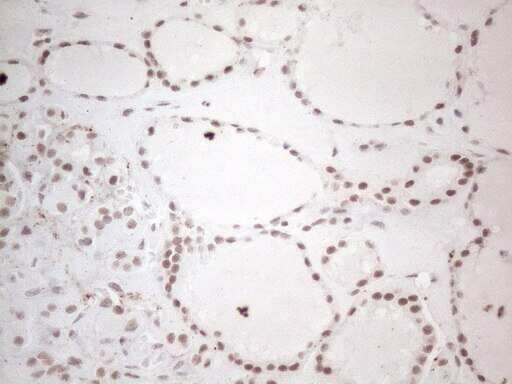
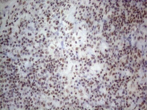
Supportive validation
- Submitted by
- Invitrogen Antibodies (provider)
- Main image
- Experimental details
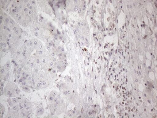
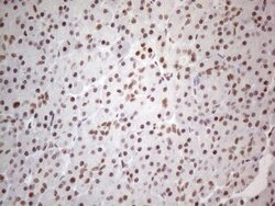
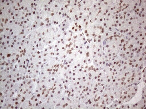
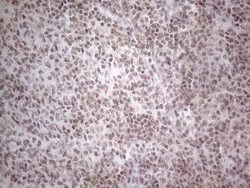
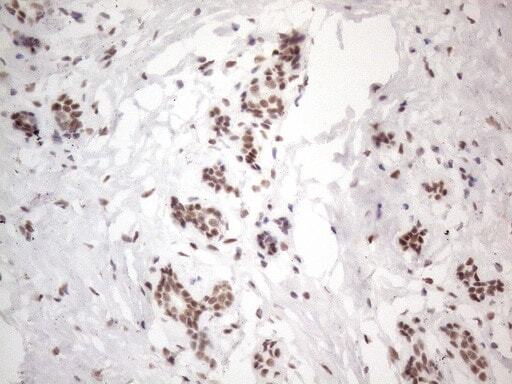
- Immunohistochemistry was performed on paraffin-embedded human thyroid tissue. To expose target proteins, heat-induced epitope retrieval by 1mM EDTA in 10mM Tris buffer (pH8.5) at 120°C for 3 min. Following antigen retrieval, tissues were probed with a NR2C2 monoclonal antibody (Product # MA5-26855) at a dilution of 1:150.
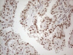
- Submitted by
- Invitrogen Antibodies (provider)
- Main image
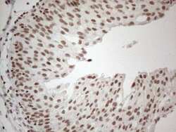
- Experimental details
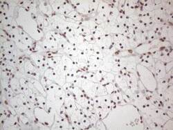
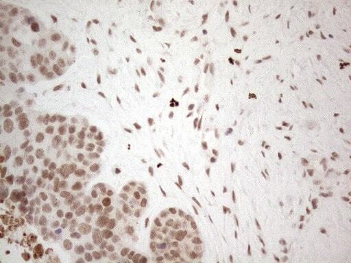
- Immunohistochemistry was performed on paraffin-embedded human bladder tissue. To expose target proteins, heat-induced epitope retrieval by 1mM EDTA in 10mM Tris buffer (pH8.5) at 120°C for 3 min. Following antigen retrieval, tissues were probed with a NR2C2 monoclonal antibody (Product # MA5-26855) at a dilution of 1:150.
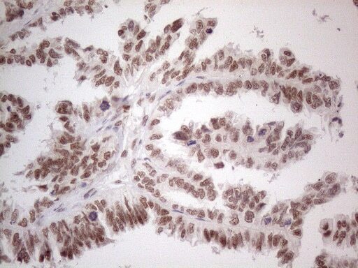
- Submitted by
- Invitrogen Antibodies (provider)
- Main image

- Experimental details
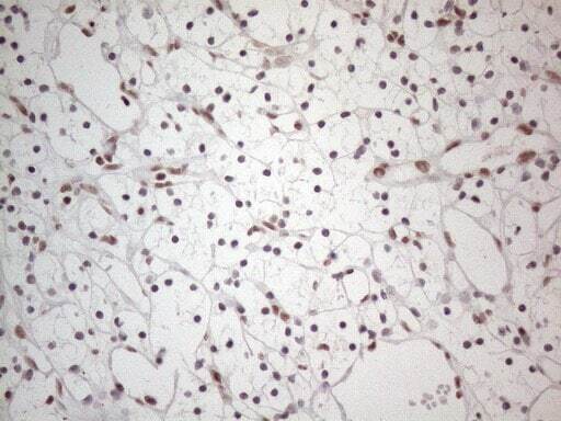
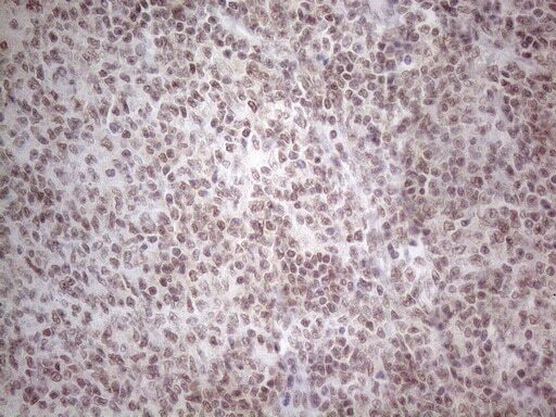
- Immunohistochemistry was performed on paraffin-embedded human lymph node tissue. To expose target proteins, heat-induced epitope retrieval by 1mM EDTA in 10mM Tris buffer (pH8.5) at 120°C for 3 min. Following antigen retrieval, tissues were probed with a NR2C2 monoclonal antibody (Product # MA5-26855) at a dilution of 1:150.
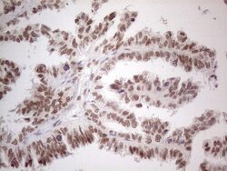
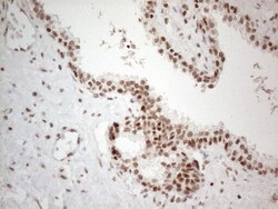
- Submitted by
- Invitrogen Antibodies (provider)
- Main image
- Experimental details
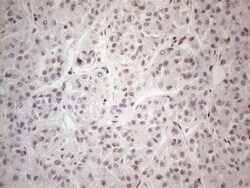
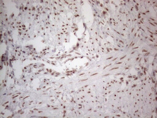
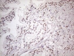
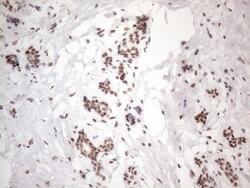
- Immunohistochemistry was performed on paraffin-embedded carcinoma of human thyroid tissue. To expose target proteins, heat-induced epitope retrieval by 1mM EDTA in 10mM Tris buffer (pH8.5) at 120°C for 3 min. Following antigen retrieval, tissues were probed with a NR2C2 monoclonal antibody (Product # MA5-26855) at a dilution of 1:150.

- Submitted by
- Invitrogen Antibodies (provider)
- Main image
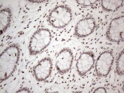
- Experimental details
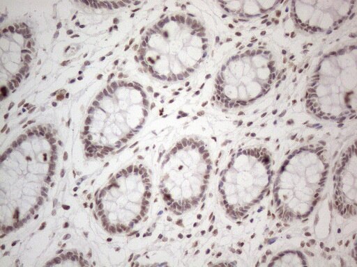
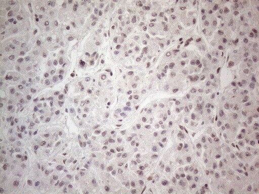
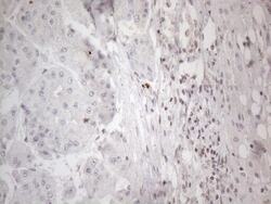
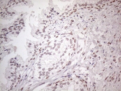
- Immunohistochemistry was performed on paraffin-embedded human colon tissue. To expose target proteins, heat-induced epitope retrieval by 1mM EDTA in 10mM Tris buffer (pH8.5) at 120°C for 3 min. Following antigen retrieval, tissues were probed with a NR2C2 monoclonal antibody (Product # MA5-26855) at a dilution of 1:150.
- Submitted by
- Invitrogen Antibodies (provider)
- Main image
- Experimental details
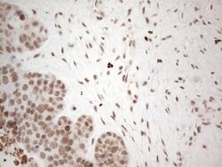
- Immunohistochemistry was performed on paraffin-embedded adenocarcinoma of human colon tissue. To expose target proteins, heat-induced epitope retrieval by 1mM EDTA in 10mM Tris buffer (pH8.5) at 120°C for 3 min. Following antigen retrieval, tissues were probed with a NR2C2 monoclonal antibody (Product # MA5-26855) at a dilution of 1:150.
- Submitted by
- Invitrogen Antibodies (provider)
- Main image
- Experimental details
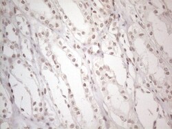
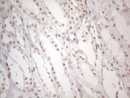
- Immunohistochemistry was performed on paraffin-embedded human kidney tissue. To expose target proteins, heat-induced epitope retrieval by 1mM EDTA in 10mM Tris buffer (pH8.5) at 120°C for 3 min. Following antigen retrieval, tissues were probed with a NR2C2 monoclonal antibody (Product # MA5-26855) at a dilution of 1:150.
- Submitted by
- Invitrogen Antibodies (provider)
- Main image
- Experimental details
- Immunohistochemistry was performed on paraffin-embedded human liver tissue. To expose target proteins, heat-induced epitope retrieval by 1mM EDTA in 10mM Tris buffer (pH8.5) at 120°C for 3 min. Following antigen retrieval, tissues were probed with a NR2C2 monoclonal antibody (Product # MA5-26855) at a dilution of 1:150.
- Submitted by
- Invitrogen Antibodies (provider)
- Main image
- Experimental details
- Immunohistochemistry was performed on paraffin-embedded human ovary tissue. To expose target proteins, heat-induced epitope retrieval by 1mM EDTA in 10mM Tris buffer (pH8.5) at 120°C for 3 min. Following antigen retrieval, tissues were probed with a NR2C2 monoclonal antibody (Product # MA5-26855) at a dilution of 1:150.
- Submitted by
- Invitrogen Antibodies (provider)
- Main image
- Experimental details
- Immunohistochemistry was performed on paraffin-embedded adenocarcinoma of human ovary tissue. To expose target proteins, heat-induced epitope retrieval by 1mM EDTA in 10mM Tris buffer (pH8.5) at 120°C for 3 min. Following antigen retrieval, tissues were probed with a NR2C2 monoclonal antibody (Product # MA5-26855) at a dilution of 1:150.
- Submitted by
- Invitrogen Antibodies (provider)
- Main image
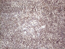
- Experimental details
- Immunohistochemistry was performed on paraffin-embedded human tonsil tissue. To expose target proteins, heat-induced epitope retrieval by 1mM EDTA in 10mM Tris buffer (pH8.5) at 120°C for 3 min. Following antigen retrieval, tissues were probed with a NR2C2 monoclonal antibody (Product # MA5-26855) at a dilution of 1:150.
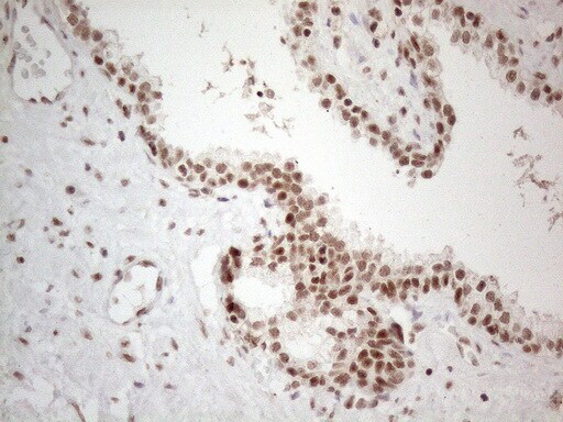
- Submitted by
- Invitrogen Antibodies (provider)
- Main image
- Experimental details
- Immunohistochemistry was performed on paraffin-embedded carcinoma of human prostate tissue. To expose target proteins, heat-induced epitope retrieval by 1mM EDTA in 10mM Tris buffer (pH8.5) at 120°C for 3 min. Following antigen retrieval, tissues were probed with a NR2C2 monoclonal antibody (Product # MA5-26855) at a dilution of 1:150.
- Submitted by
- Invitrogen Antibodies (provider)
- Main image
- Experimental details
- Immunohistochemistry was performed on paraffin-embedded human endometrium tissue. To expose target proteins, heat-induced epitope retrieval by 1mM EDTA in 10mM Tris buffer (pH8.5) at 120°C for 3 min. Following antigen retrieval, tissues were probed with a NR2C2 monoclonal antibody (Product # MA5-26855) at a dilution of 1:150.
- Submitted by
- Invitrogen Antibodies (provider)
- Main image
- Experimental details
- Immunohistochemistry was performed on paraffin-embedded carcinoma of human kidney tissue. To expose target proteins, heat-induced epitope retrieval by 1mM EDTA in 10mM Tris buffer (pH8.5) at 120°C for 3 min. Following antigen retrieval, tissues were probed with a NR2C2 monoclonal antibody (Product # MA5-26855) at a dilution of 1:150.
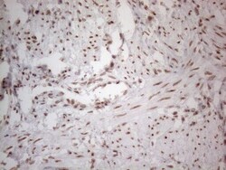
- Submitted by
- Invitrogen Antibodies (provider)
- Main image
- Experimental details
- Immunohistochemistry was performed on paraffin-embedded carcinoma of human liver tissue. To expose target proteins, heat-induced epitope retrieval by 1mM EDTA in 10mM Tris buffer (pH8.5) at 120°C for 3 min. Following antigen retrieval, tissues were probed with a NR2C2 monoclonal antibody (Product # MA5-26855) at a dilution of 1:150.
- Submitted by
- Invitrogen Antibodies (provider)
- Main image
- Experimental details
- Immunohistochemistry was performed on paraffin-embedded human pancreas tissue. To expose target proteins, heat-induced epitope retrieval by 1mM EDTA in 10mM Tris buffer (pH8.5) at 120°C for 3 min. Following antigen retrieval, tissues were probed with a NR2C2 monoclonal antibody (Product # MA5-26855) at a dilution of 1:150.
- Submitted by
- Invitrogen Antibodies (provider)
- Main image
- Experimental details
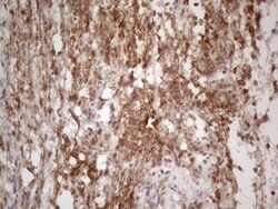
- Immunohistochemistry was performed on paraffin-embedded human lymphoma tissue. To expose target proteins, heat-induced epitope retrieval by 1mM EDTA in 10mM Tris buffer (pH8.5) at 120°C for 3 min. Following antigen retrieval, tissues were probed with a NR2C2 monoclonal antibody (Product # MA5-26855) at a dilution of 1:150.
- Submitted by
- Invitrogen Antibodies (provider)
- Main image
- Experimental details
- Immunohistochemistry was performed on paraffin-embedded human prostate tissue. To expose target proteins, heat-induced epitope retrieval by 1mM EDTA in 10mM Tris buffer (pH8.5) at 120°C for 3 min. Following antigen retrieval, tissues were probed with a NR2C2 monoclonal antibody (Product # MA5-26855) at a dilution of 1:150.
- Submitted by
- Invitrogen Antibodies (provider)
- Main image
- Experimental details
- Immunohistochemistry was performed on paraffin-embedded adenocarcinoma of human endometrium tissue. To expose target proteins, heat-induced epitope retrieval by 1mM EDTA in 10mM Tris buffer (pH8.5) at 120°C for 3 min. Following antigen retrieval, tissues were probed with a NR2C2 monoclonal antibody (Product # MA5-26855) at a dilution of 1:150.
- Submitted by
- Invitrogen Antibodies (provider)
- Main image
- Experimental details
- Immunohistochemistry was performed on paraffin-embedded human breast tissue. To expose target proteins, heat-induced epitope retrieval by 1mM EDTA in 10mM Tris buffer (pH8.5) at 120°C for 3 min. Following antigen retrieval, tissues were probed with a NR2C2 monoclonal antibody (Product # MA5-26855) at a dilution of 1:150.
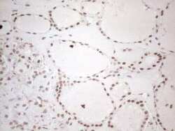
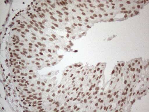
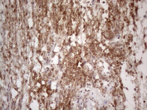
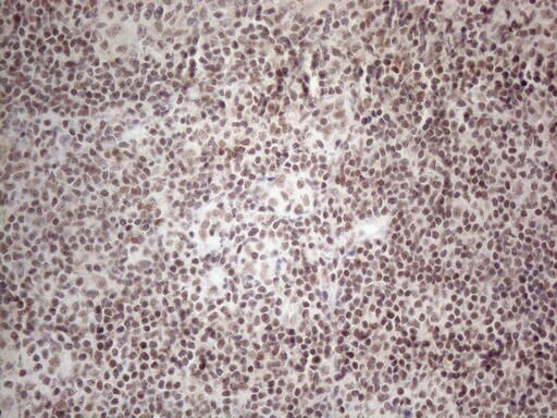
Supportive validation
- Submitted by
- Invitrogen Antibodies (provider)
- Main image
- Experimental details
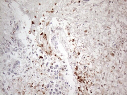
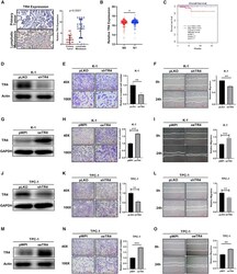
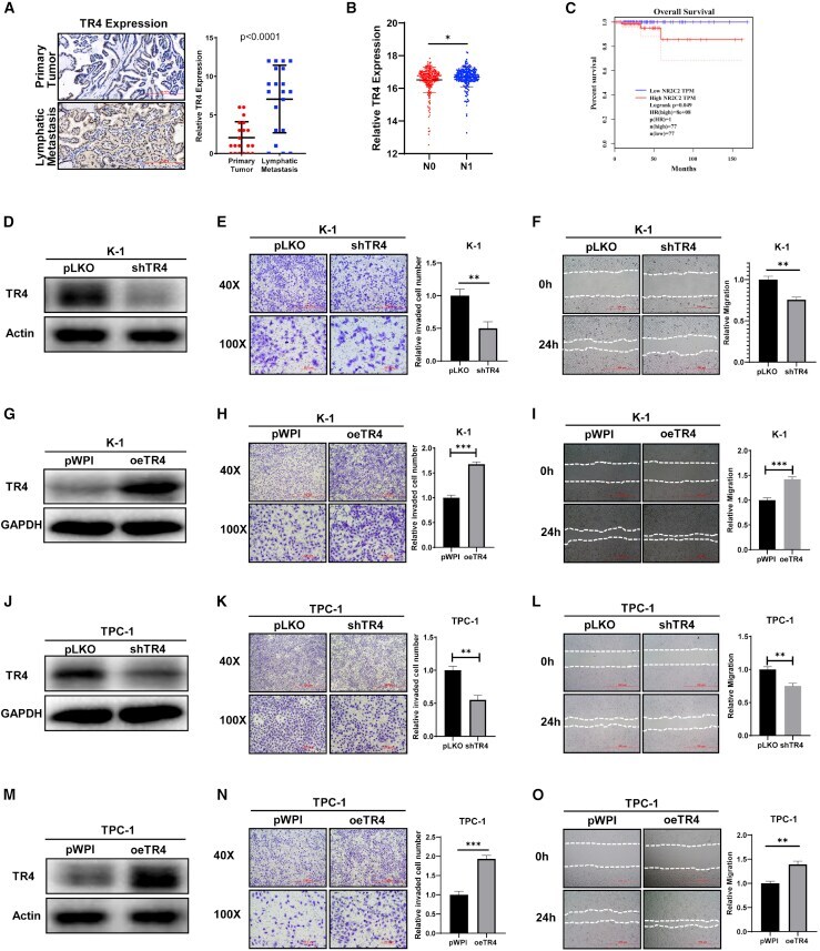
- Figure 1 TR4 promotes cell invasion/migration of PTC cells (A) TR4 expression level was examined in 20 pairs of primary PTC tumors and the paired lymph node metastatic PTC tumors using IHC staining strategy. Left, representative images of TR4 signal in PTC patients; top, image from primary PTC tumor; bottom, image from the paired PTC tumors metastasizing into lymph node; right, quantification of TR4 signal in PTC patients. Paired t test was used to make the statistical analysis. (B) TR4 expression level was significantly increased in nodal metastasis (N1) TC patients compared to non-metastasis (N0) TC patients. (C) OS analysis of TC patients classified by TR4 expression level. The blue line stands for low TR4 TPM; red line stands for high TR4 TPM. (D) Western blot assay was used to check the efficiency of TR4 knockdown in K-1 cells. Actin was internal control. (E) Knockdown of TR4 suppressed the cell invasion of K-1 cells. Left, representative images of invading K-1 cells; right, statistical analysis of invading K-1 cells. (F) Wound-healing assay showed that knockdown of TR4 inhibited cell migration of K-1 cells. Left, representative images of migrating K-1 cells; right, statistical analysis of migrating K-1 cells. (G) Western blot assay was used to check the efficiency of TR4 overexpression (oeTR4) in K-1 cells. GAPDH was loading control. (H) oeTR4 promoted cell invasion of K-1 cells. Left, representative images of invading K-1 cells; right, statistical analysis of invading
- Submitted by
- Invitrogen Antibodies (provider)
- Main image

- Experimental details
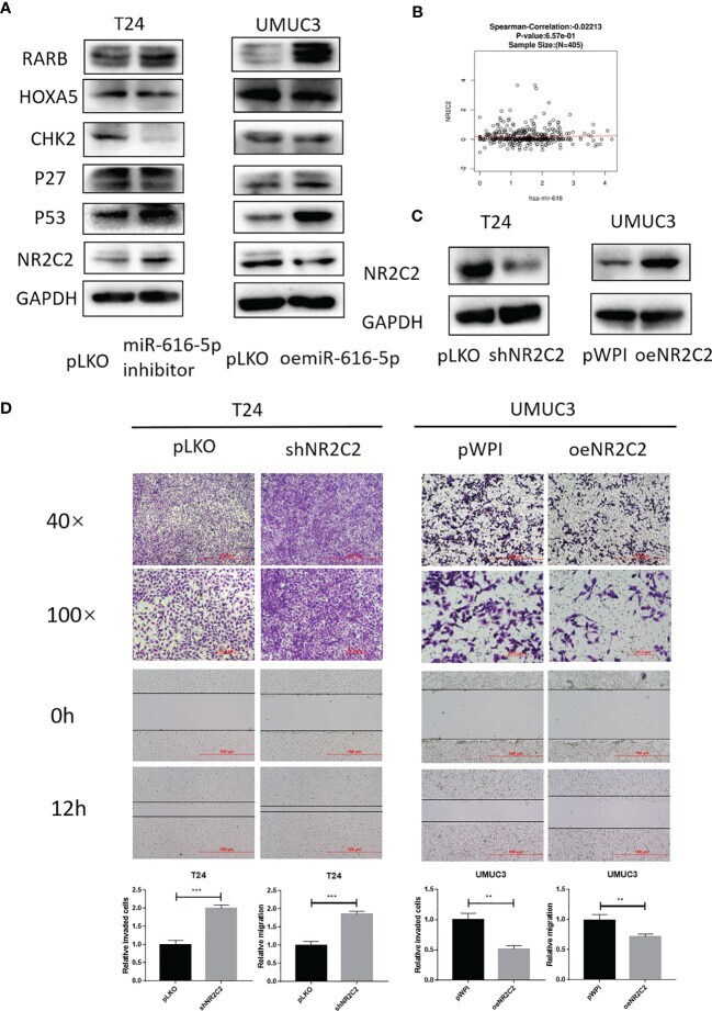
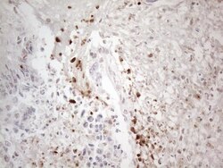
- Figure 3 NR2C2 is negatively correlated with miR-616-5p and inhibits bladder cancer cell invasion and migration. (A) Western blot assay for screening the downstream target genes in T24 cells transduced with pLKO and miR-616-5p inhibitor and UMUC3 cells transduced with pLKO and oemiR-616-5p. (B) Correlation between miR-616 and NR2C2 expression in bladder cancer according to LinkedOmics database. (C) Western blot assay for NR2C2 expression in T24 cells transduced with pLKO and shNR2C2 and UMUC3 cells transduced with pWPI and oeNR2C2. (D) Chamber-transwell invasion and wound healing migration assays performed in T24 cells transduced with pLKO and shNR2C2 and UMUC3 cells transduced with pWPI and oeNR2C2. For (C) , quantification data are presented as mean +- SD, and significant differences are indicated by ** P < 0.01, and *** P < 0.001 compared to the controls.