 Explore
 Validate
 Learn
Learn Western blot
Antibody data
- Antibody Data
- Antigen structure
- References [0]
- Comments [0]
- Validations
- Western blot [1]
- Immunohistochemistry [1]
Submit
Validation data
Reference
Comment
Report error
- Product number
- AF6820 - Provider product page

- Provider
- R&D Systems
- Product name
- Mouse/Rat Testisin/Prss21 Antibody
- Antibody type
- Polyclonal
- Description
- Immunogen affinity purified. Detects recombinant mouse Testisin/Prss21 in direct ELISAs and Western blots. Detects mouse and rat Testisin/Prss21 in Western blots. In direct ELISAs, less than 1% cross-reactivity with recombinant mouse (rm) Prss8 and rmPrss27 is observed.
- Reactivity
- Mouse, Rat
- Host
- Goat
- Conjugate
- Unconjugated
- Antigen sequence
Q9JHJ7- Isotype
- IgG
- Vial size
- 100 ug
- Concentration
- LYOPH
- Storage
- Use a manual defrost freezer and avoid repeated freeze-thaw cycles. 12 months from date of receipt, -20 to -70 °C as supplied. 1 month, 2 to 8 °C under sterile conditions after reconstitution. 6 months, -20 to -70 °C under sterile conditions after reconstitution.
No comments: Submit comment
Supportive validation
- Submitted by
- R&D Systems (provider)
- Main image

- Experimental details
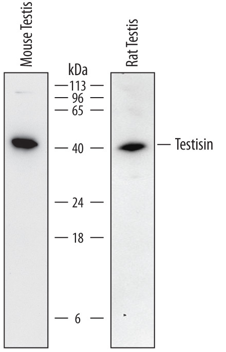
- Detection of Mouse and Rat Testisin/Prss21 by Western Blot. Western blot shows lysates of mouse testis tissue and rat testis tissue. PVDF membrane was probed with 0.5 µg/mL of Goat Anti-Mouse Testisin/Prss21 Antigen Affinity-purified Polyclonal Antibody (Catalog # AF6820) followed by HRP-conjugated Anti-Goat IgG Secondary Antibody (Catalog # HAF017). A specific band was detected for Testisin/Prss21 at approximately 40 kDa (as indicated). This experiment was conducted under reducing conditions and using Immunoblot Buffer Group 1.
Supportive validation
- Submitted by
- R&D Systems (provider)
- Main image
- Experimental details
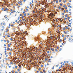
- Testisin/Prss21 in Mouse Testis. Testisin/Prss21 was detected in perfusion fixed frozen sections of mouse testis using Goat Anti-Mouse Testisin/Prss21 Antigen Affinity-purified Polyclonal Antibody (Catalog # AF6820) at 1.7 µg/mL overnight at 4 °C. Tissue was stained using the Anti-Goat HRP-DAB Cell & Tissue Staining Kit (brown; Catalog # CTS008) and counterstained with hematoxylin (blue). Specific staining was localized to the plasma membranes of spermatocytes. View our protocol for Chromogenic IHC Staining of Frozen Tissue Sections. This application has not been tested in rat samples.
