 Explore
 Validate
 Learn
Learn Western blot
 Immunocytochemistry
Antibody data
- Antibody Data
- Antigen structure
- References [3]
- Comments [0]
- Validations
- Western blot [3]
- Immunohistochemistry [5]
Submit
Validation data
Reference
Comment
Report error
- Product number
- NBP1-89557 - Provider product page

- Provider
- Novus Biologicals
- Proper citation
- Novus Cat#NBP1-89557, RRID:AB_11026380
- Product name
- Rabbit Polyclonal CLPP Antibody
- Antibody type
- Polyclonal
- Description
- Immunogen affinity purified. Specificity of human CLPP antibody verified on a Protein Array containing target protein plus 383 other non-specific proteins.
- Reactivity
- Human, Mouse, Rat
- Host
- Rabbit
- Isotype
- IgG
- Vial size
- 0.1 ml
- Storage
- Store at 4C short term. Aliquot and store at -20C long term. Avoid freeze-thaw cycles.
Submitted references Alpha-synuclein suppresses mitochondrial protease ClpP to trigger mitochondrial oxidative damage and neurotoxicity.
Perrault syndrome is caused by recessive mutations in CLPP, encoding a mitochondrial ATP-dependent chambered protease.
Systematic validation of antibody binding and protein subcellular localization using siRNA and confocal microscopy.
Hu D, Sun X, Liao X, Zhang X, Zarabi S, Schimmer A, Hong Y, Ford C, Luo Y, Qi X
Acta neuropathologica 2019 Jun;137(6):939-960
Acta neuropathologica 2019 Jun;137(6):939-960
Perrault syndrome is caused by recessive mutations in CLPP, encoding a mitochondrial ATP-dependent chambered protease.
Jenkinson EM, Rehman AU, Walsh T, Clayton-Smith J, Lee K, Morell RJ, Drummond MC, Khan SN, Naeem MA, Rauf B, Billington N, Schultz JM, Urquhart JE, Lee MK, Berry A, Hanley NA, Mehta S, Cilliers D, Clayton PE, Kingston H, Smith MJ, Warner TT, University of Washington Center for Mendelian Genomics., Black GC, Trump D, Davis JR, Ahmad W, Leal SM, Riazuddin S, King MC, Friedman TB, Newman WG
American journal of human genetics 2013 Apr 4;92(4):605-13
American journal of human genetics 2013 Apr 4;92(4):605-13
Systematic validation of antibody binding and protein subcellular localization using siRNA and confocal microscopy.
Stadler C, Hjelmare M, Neumann B, Jonasson K, Pepperkok R, Uhlén M, Lundberg E
Journal of proteomics 2012 Apr 3;75(7):2236-51
Journal of proteomics 2012 Apr 3;75(7):2236-51
No comments: Submit comment
Supportive validation
- Submitted by
- Novus Biologicals (provider)
- Main image

- Experimental details
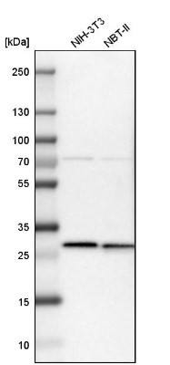
- Western Blot: CLPP Antibody [NBP1-89557] - Analysis in mouse cell line NIH-3T3 and rat cell line NBT-II.
- Submitted by
- Novus Biologicals (provider)
- Main image
- Experimental details
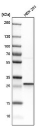
- Western Blot: CLPP Antibody [NBP1-89557] - Analysis in human cell line HEK 293.

- Submitted by
- Novus Biologicals (provider)
- Main image

- Experimental details
- Western Blot: CLPP Antibody [NBP1-89557] - Analysis in HEK293 cells transfected with control siRNA, target specific siRNA probe #1, using Anti-CLPP antibody. Remaining relative intensity is presented. Loading control: Anti-PPIB.

Supportive validation
- Submitted by
- Novus Biologicals (provider)
- Main image

- Experimental details
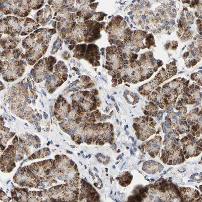
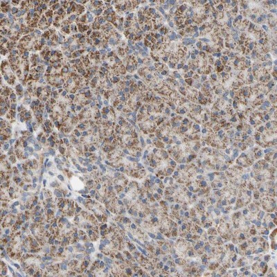
- Immunohistochemistry-Paraffin: CLPP Antibody [NBP1-89557] - Staining of human pancreas shows strong granular cytoplasmic positivity in exocrine glandular cells.
- Submitted by
- Novus Biologicals (provider)
- Main image
- Experimental details
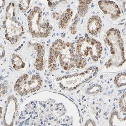
- Immunohistochemistry-Paraffin: CLPP Antibody [NBP1-89557] - Staining of human kidney shows moderate granular cytoplasmic positivity in cells in tubules.

- Submitted by
- Novus Biologicals (provider)
- Main image
- Experimental details
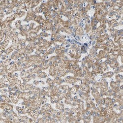
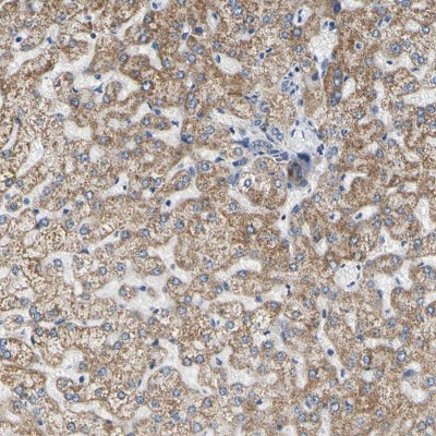
- Immunohistochemistry-Paraffin: CLPP Antibody [NBP1-89557] - Staining of human liver shows moderate granular cytoplasmic positivity in hepatocytes.
- Submitted by
- Novus Biologicals (provider)
- Main image
- Experimental details
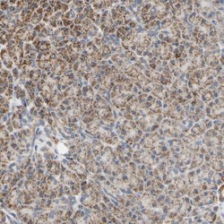
- Immunohistochemistry-Paraffin: CLPP Antibody [NBP1-89557] - Staining of human pancreas shows moderate granular cytoplasmic positivity in exocrine glandular cells.
- Submitted by
- Novus Biologicals (provider)
- Main image
- Experimental details
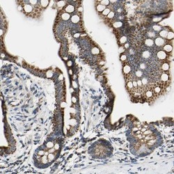
- Immunohistochemistry-Paraffin: CLPP Antibody [NBP1-89557] - Staining of human small intestine shows strong granular cytoplasmic positivity in glandular cells.
