Explore
 Validate
 Learn
LearnAPC-045-200UL
antibody from Invitrogen Antibodies
Targeting: CNGA2
CNCA, CNCA1, CNG2, FLJ46312, OCNC1, OCNCa, OCNCALPHA
 Western blot
Antibody data
- Antibody Data
- Antigen structure
- References [0]
- Comments [0]
- Validations
- Western blot [2]
- Immunocytochemistry [1]
- Immunohistochemistry [2]
Submit
Validation data
Reference
Comment
Report error
- Product number
- APC-045-200UL - Provider product page

- Provider
- Invitrogen Antibodies
- Product name
- CNGA2 Polyclonal Antibody
- Antibody type
- Polyclonal
- Antigen
- Other
- Reactivity
- Human, Mouse, Rat
- Host
- Rabbit
- Isotype
- IgG
- Vial size
- 200 µL
- Concentration
- 0.8 mg/mL
- Storage
- -20° C, Avoid Freeze/Thaw Cycles
No comments: Submit comment
Supportive validation
- Submitted by
- Invitrogen Antibodies (provider)
- Main image
- Experimental details
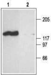
- Western blot analysis of rat brain membranes: - 1. Anti-CNGA2 Antibody (#APC-045), (1:200). 2. Anti-CNGA2 Antibody , preincubated with CNGA2 Blocking Peptide (#BLP-PC045).
- Submitted by
- Invitrogen Antibodies (provider)
- Main image

- Experimental details
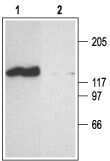
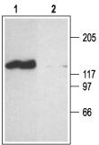
- Western blot analysis of rat brain membranes: - 1. Anti-CNGA2 Antibody (#APC-045), (1:200). 2. Anti-CNGA2 Antibody , preincubated with CNGA2 Blocking Peptide (#BLP-PC045).
Supportive validation
- Submitted by
- Invitrogen Antibodies (provider)
- Main image

- Experimental details
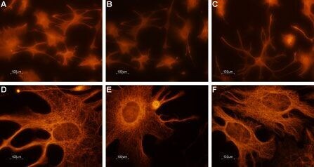
- Expression of CNGA2 in rat cerebellum primary culture - Immunocytochemical staining of paraformaldehyde-fixed and permeabilized rat cerebellum primary culture. A-F. Immunocytochemical staining using Anti-CNGA2 Antibody (#APC-045), (1:100) followed by goat Anti-rabbit-AlexaFluor-555secondary Antibody . Magnification:A-C: x20D-F: x100.
Supportive validation
- Submitted by
- Invitrogen Antibodies (provider)
- Main image
- Experimental details
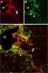
- Expression of CNGA2 in mouse cerebrum - Immunohistochemical staining of mouse cerebrum using Anti-CNGA2 Antibody (#APC-045). A. CNGA2 (red) appears in cells lining up the wall of the lateral ventricle (LV) (horizontal arrows). B. Staining of astrocytes with mouse Anti-glial fibrillary acidic protein (GFAP, green) demonstrates penetration of astrocytic fibers (vertical arrows) into the wall of the lateral ventricle. C. Confocal merge of panels A and B.

- Submitted by
- Invitrogen Antibodies (provider)
- Main image

- Experimental details
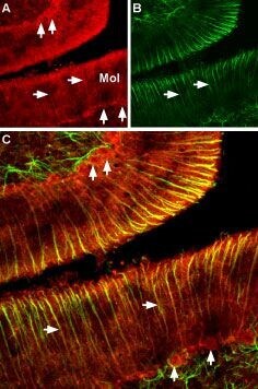
- Expression of CNGA2 in rat cerebellum - Immunohistochemical staining of rat cerebellum using Anti-CNGA2 Antibody (#APC-045). A. CNGA2 (red) appears in Purkinje cells (vertical arrows) and in astrocytic fibers (horizontal arrows) traversing the cerebellar molecular layer (Mol). B. Staining of astrocytes with mouse Anti-glial fibrillary acidic protein (GFAP, green) demonstrates the full distribution of astrocytic fibers (horizontal arrows) in the cerebellum. C. Confocal merge of panels A and B.