 Explore
 Validate
 Learn
Learn Western blot
 Other assay
Antibody data
- Antibody Data
- Antigen structure
- References [5]
- Comments [0]
- Validations
- Other assay [2]
Submit
Validation data
Reference
Comment
Report error
- Product number
- PA1-960 - Provider product page

- Provider
- Invitrogen Antibodies
- Product name
- PSME1 Polyclonal Antibody
- Antibody type
- Polyclonal
- Antigen
- Synthetic peptide
- Description
- PA1-960 detects proteasome activator 11S REG alpha subunit from canine, hamster, human, mouse and rat tissues and cells. PA1-960 has been successfully used in Western blot procedures. By Western blot, this antibody detects a 28 kDa protein representing 11S REG alpha subunit in rat brain extract. PA1-960 immunizing peptide corresponds to amino acid residues 5-22 of human proteasome activator 11S REG alpha. This sequence differs from the mouse and rat 11S REG alpha protein by a single amino acid residue. PA1-960 immunizing peptide (Cat. # PEP-097) is available for use in neutralization and control experiments.
- Reactivity
- Human, Mouse, Rat, Canine, Hamster
- Host
- Rabbit
- Isotype
- IgG
- Vial size
- 100 µg
- Concentration
- 1 mg/mL
- Storage
- -20° C, Avoid Freeze/Thaw Cycles
Submitted references Heterogeneous nuclear ribonucleoprotein A2 participates in the replication of Japanese encephalitis virus through an interaction with viral proteins and RNA.
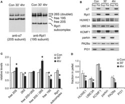
Characterization of the Brain 26S Proteasome and its Interacting Proteins.
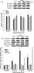
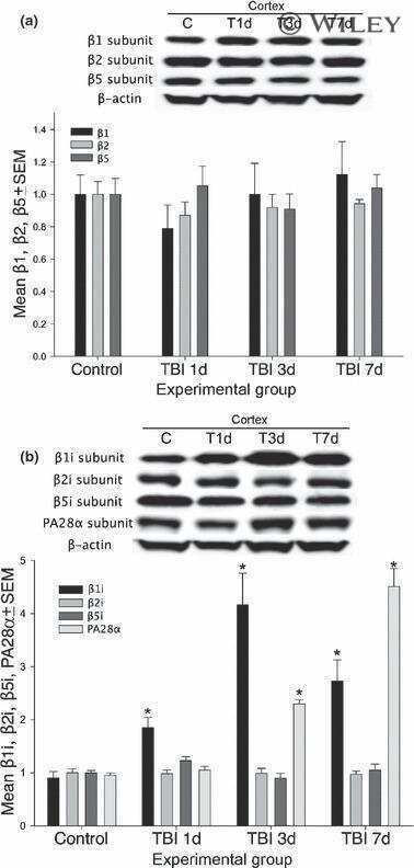
Alterations of cerebral cortex and hippocampal proteasome subunit expression and function in a traumatic brain injury rat model.
Reduced ubiquitin C-terminal hydrolase-1 expression levels in dementia with Lewy bodies.
Proteasome impairment does not contribute to pathogenesis in R6/2 Huntington's disease mice: exclusion of proteasome activator REGgamma as a therapeutic target.
Katoh H, Mori Y, Kambara H, Abe T, Fukuhara T, Morita E, Moriishi K, Kamitani W, Matsuura Y
Journal of virology 2011 Nov;85(21):10976-88
Journal of virology 2011 Nov;85(21):10976-88
Characterization of the Brain 26S Proteasome and its Interacting Proteins.
Tai HC, Besche H, Goldberg AL, Schuman EM
Frontiers in molecular neuroscience 2010;3
Frontiers in molecular neuroscience 2010;3
Alterations of cerebral cortex and hippocampal proteasome subunit expression and function in a traumatic brain injury rat model.
Yao X, Liu J, McCabe JT
Journal of neurochemistry 2008 Jan;104(2):353-63
Journal of neurochemistry 2008 Jan;104(2):353-63
Reduced ubiquitin C-terminal hydrolase-1 expression levels in dementia with Lewy bodies.
Barrachina M, Castaño E, Dalfó E, Maes T, Buesa C, Ferrer I
Neurobiology of disease 2006 May;22(2):265-73
Neurobiology of disease 2006 May;22(2):265-73
Proteasome impairment does not contribute to pathogenesis in R6/2 Huntington's disease mice: exclusion of proteasome activator REGgamma as a therapeutic target.
Bett JS, Goellner GM, Woodman B, Pratt G, Rechsteiner M, Bates GP
Human molecular genetics 2006 Jan 1;15(1):33-44
Human molecular genetics 2006 Jan 1;15(1):33-44
No comments: Submit comment
Supportive validation
- Submitted by
- Invitrogen Antibodies (provider)
- Main image
- Experimental details
- NULL
- Submitted by
- Invitrogen Antibodies (provider)
- Main image
- Experimental details
- Figure 7 Changes in proteasome complexes after NMDA exposure . (A) The disassembly of 26S proteasomes. Neuronal lysates collected at 30' and 4 h post-NMDA (20 muM, 3 min) were resolved by 2-5% gradient native gel and immunoblotted against 20S subunit alpha7 and 19S subunit Rpt1. (B) Changes in proteasome-associated proteins. Lysates from (A) were subjected to ultracentrifugation to sediment proteasomes. Equal amounts of supernatant (Sup) and pellet (Pel) materials were analyzed by SDS-PAGE. The sedimentation property of proteasome-interacting proteins (HUWE1, UBE3A, KCMF1, PA28alpha) was examined by Western blotting. Parkin and PI31 were not detected in the pellet. (C) Quantification of changes in proteasome distributions in (A) by densitometry ( n = 6, mean +- SEM, * p < 0.05 by paired t -test). (D) The graph represents the sedimentation data in (B) . The protein level in the pellet is divided by the total level (supernatant + pellet), and the results are plotted ( n = 4, * p < 0.05 by paired t -test).
