Explore
 Validate
 Learn
Learn Western blot
 Immunohistochemistry
Antibody data
- Antibody Data
- Antigen structure
- References [1]
- Comments [0]
- Validations
- Immunohistochemistry [1]
- Other assay [1]
Submit
Validation data
Reference
Comment
Report error
- Product number
- PA1-84183 - Provider product page

- Provider
- Invitrogen Antibodies
- Product name
- IL-17A Polyclonal Antibody
- Antibody type
- Polyclonal
- Antigen
- Recombinant full-length protein
- Description
- Heat-mediated antigen retrieval using a sodium citrate buffer (pH 6.0) is recommended for the staining of paraffin sections. This antibody may be used in Western Blotting applications under either reducing or non-reducing conditions. Reconstitute with 0.1 mL of distilled water. For long term storage the addition of 0.09% sodium azide is recommended (unless the product will be used for functional studies). Prior to reconstitution, store at 4ºC. After reconstitution, store undiluted at -20ºC, avoiding freeze/thaw cycles. Rabbit anti Human Interleukin-17 antibody recognizes human IL-17 (Interleukin-17/17A), also known as Cytotoxic T-lymphocyte-associated antigen 8 or CTLA-8.
- Reactivity
- Human
- Host
- Rabbit
- Isotype
- IgG
- Vial size
- 100 µg
- Concentration
- 1 mg/mL
- Storage
- -20°C
Submitted references Increased Serum Level and High Tissue Immunoexpression of Interleukin 17 in Cutaneous Lichen Planus: A Novel Therapeutic Target for Recalcitrant Cases?
Żychowska M, Batycka-Baran A, Baran W
Disease markers 2020;2020:6521274
Disease markers 2020;2020:6521274
No comments: Submit comment
Supportive validation
- Submitted by
- Invitrogen Antibodies (provider)
- Main image

- Experimental details
- Immunohistochemical analysis of IL-17A in formalin fixed,paraffin embedded human breast invasive ductal carcinoma. Samples were incubated in IL-17A Polyclonal antibody (Product # PA1-84183).

Supportive validation
- Submitted by
- Invitrogen Antibodies (provider)
- Main image

- Experimental details
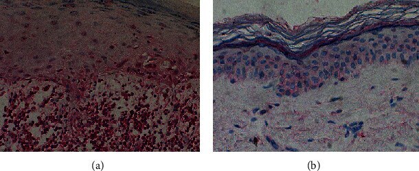
- Figure 2 Representative immunohistochemical stainings of IL-17 expression: (a) high number of cells showing positive staining in CLP lesions and (b) single cells showing positive staining in a healthy skin. Magnification, x200.