 Explore
 Validate
 Learn
Learn Western blot
Antibody data
- Antibody Data
- Antigen structure
- References [0]
- Comments [0]
- Validations
- Western blot [2]
- Immunocytochemistry [1]
- Immunohistochemistry [1]
- Flow cytometry [1]
Submit
Validation data
Reference
Comment
Report error
- Product number
- ABIN2508492 - Provider product page

- Provider
- antibodies-online
- Product name
- anti-Interleukin 17A (IL17A) antibody (Biotin)
- Antibody type
- Polyclonal
- Antigen
- Other
- Description
- Produced from sera of rabbits pre-immunized with highly pure (>98%) recombinant hIL-17A. Anti-Human IL-17A specific antibody was purified by affinity chromatography and then biotinylated.
- Reactivity
- Human
- Host
- Rabbit
- Conjugate
- Biotin
- Vial size
- 50 μg
- Storage
- -20°C
No comments: Submit comment
Supportive validation
- Submitted by
- antibodies-online (provider)
- Main image
- Experimental details
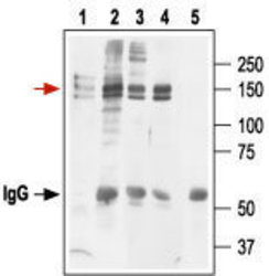
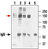
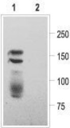
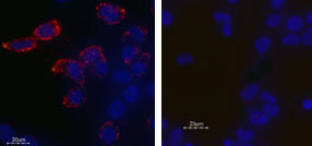
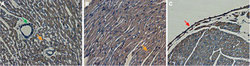
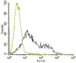
- Western blot analysis of Kv11.1 transfected HEK-293 cells: 1. Anti-Kv11.1 (HERG) (extracellular) antibody (ABIN2511224), (1:200). 2. Anti-Kv11.1 (HERG) (extracellular) antibody, preincubated with the control peptide antigen. Expression of Kv11.1 in rat heart Immunohistochemical staining of rat heart using Anti-Kv11.1 (HERG) (extracellular) antibody (ABIN2511224). A. Transversal section of the atrium wall, note that arterial smooth muscle fibers were not stained (green arrow). B. Longitudinal section of the myocardium. C. Section showing myocardium and endocardium (red arrow). DAB product is brown and the counterstain is cresyl violet. Expression of Kv11.1 in HEK-293 transfected cells Immunocytochemical staining of live intact HEK-293 transfected cells.Cells were stained with Anti-Kv11.1 (HERG) (extracellular) antibody (ABIN2511224), (1:25) (left panel) or with Kv11.1 (HERG) (extracellular) antibody preincubated with the control peptide antigen (right panel), followed by goat anti-rabbit-AlexaFluor-555 secondary antibody (red). Nuclei of the living cells were stained with the cell permeable dye Hoechst 33342 (blue). Note that not all cells are stained, indicating that not all the Kv11.1 channels are expressed at the cell membrane at the moment of staining. Indirect flow cytometry analysis of human chronic myelogenous leukemia (K562) cells: ___ Unstained cells + goat-anti-rabbit-FITC. Black Cells + Anti-KV11.1 (HERG) (extracellular) antibody (ABIN2511224) + goat-anti-rabbit-FITC. Immunoprecipitation of Kv11.1 expressing HEK-293 cells: Cell lysate. Lysate + protein A beads + Anti-Kv11.1 (HERG) (extracellular) antibody (ABIN2511224). Lysate + protein A beads + Anti-Kv11.1 (erg1) antibody (APC-016). Lysate + protein A beads + Anti-hKv11.1 (HERG) antibody (APC-062). Lysate + protein A beads + pre-immune rabbit serum. Red arrow indicates Kv11.1 while the black arrow shows the IgG heavy chain. Immunoblot was performed with Anti-Kv11.1 (HERG) (extracellular) antibody.
- Submitted by
- antibodies-online (provider)
- Main image
- Experimental details
- Western blot analysis of Kv11.1 transfected HEK-293 cells: 1. Anti-Kv11.1 (HERG) (extracellular) antibody (ABIN2511224), (1:200). 2. Anti-Kv11.1 (HERG) (extracellular) antibody, preincubated with the control peptide antigen. Expression of Kv11.1 in rat heart Immunohistochemical staining of rat heart using Anti-Kv11.1 (HERG) (extracellular) antibody (ABIN2511224). A. Transversal section of the atrium wall, note that arterial smooth muscle fibers were not stained (green arrow). B. Longitudinal section of the myocardium. C. Section showing myocardium and endocardium (red arrow). DAB product is brown and the counterstain is cresyl violet. Expression of Kv11.1 in HEK-293 transfected cells Immunocytochemical staining of live intact HEK-293 transfected cells.Cells were stained with Anti-Kv11.1 (HERG) (extracellular) antibody (ABIN2511224), (1:25) (left panel) or with Kv11.1 (HERG) (extracellular) antibody preincubated with the control peptide antigen (right panel), followed by goat anti-rabbit-AlexaFluor-555 secondary antibody (red). Nuclei of the living cells were stained with the cell permeable dye Hoechst 33342 (blue). Note that not all cells are stained, indicating that not all the Kv11.1 channels are expressed at the cell membrane at the moment of staining. Indirect flow cytometry analysis of human chronic myelogenous leukemia (K562) cells: ___ Unstained cells + goat-anti-rabbit-FITC. Black Cells + Anti-KV11.1 (HERG) (extracellular) antibody (ABIN2511224) + goat-anti-rabbit-FITC. Immunoprecipitation of Kv11.1 expressing HEK-293 cells: Cell lysate. Lysate + protein A beads + Anti-Kv11.1 (HERG) (extracellular) antibody (ABIN2511224). Lysate + protein A beads + Anti-Kv11.1 (erg1) antibody (APC-016). Lysate + protein A beads + Anti-hKv11.1 (HERG) antibody (APC-062). Lysate + protein A beads + pre-immune rabbit serum. Red arrow indicates Kv11.1 while the black arrow shows the IgG heavy chain. Immunoblot was performed with Anti-Kv11.1 (HERG) (extracellular) antibody.

Supportive validation
- Submitted by
- antibodies-online (provider)
- Main image

- Experimental details
- Image(s): Immunofluorescence
Supportive validation
- Submitted by
- antibodies-online (provider)
- Main image
- Experimental details
- Image(s): Immunohistochemistry

Supportive validation
- Submitted by
- antibodies-online (provider)
- Main image
- Experimental details
- Image(s): Flow cytometry
