Explore
 Validate
 Learn
Learn Flow cytometry
Antibody data
- Antibody Data
- Antigen structure
- References [54]
- Comments [0]
- Validations
- Flow cytometry [1]
- Other assay [10]
Submit
Validation data
Reference
Comment
Report error
- Product number
- 12-7178-42 - Provider product page

- Provider
- Invitrogen Antibodies
- Product name
- IL-17A Monoclonal Antibody (eBio64CAP17), PE, eBioscience™
- Antibody type
- Monoclonal
- Antigen
- Other
- Description
- Description: The eBio64CAP17 antibody reacts with human IL-17A; the antibody has been reported to cross react with Rhesus monkey IL-17A, as verified by intracellular staining experiments. The eBio64CAP17 antibody is a neutralizing antibody. This antibody has been shown to have no reactivity to human IL-17F. Reactivity to other members of the IL-17 family has not been evaluated. Interleukin-17A (IL-17A) is a CD4+ T cell-derived cytokine that promotes inflammatory responses in cell lines and is elevated in rheumatoid arthritis, asthma, multiple sclerosis, psoriasis, and transplant rejection. The cDNA encoding human IL-17A was isolated from a library of CD4+ T cells; the encoded protein exhibits 72 percent amino acid identity with HVS13 , an open reading frame from a T lymphotropic Herpesvirus saimiri, and 63 percent with mouse CTLA-8 (cytotoxic T-lymphocyte associated antigen-8). Human IL-17A exists as glycosylated 20-30 kD homodimers. High levels of IL-17A homodimer are produced by activated peripheral blood CD4+ T-cells. IL-17A enhances expression of the intracellular adhesion molecule-1 (ICAM-1) in human fibroblasts. Human IL-17A also stimulates epithelial, endothelial, or fibroblastic cells to secrete IL-6, IL-8, G-CSF, and PGE2. In the presence of human IL-17A, fibroblasts can sustain the proliferation of CD34+ hematopoietic progenitors and induce maturation into neutrophils. Mouse, rat, and human IL-17A can induce IL-6 secretion in mouse stromal cells, indicating that all homologs can recognize the mouse IL-17A receptor. IL-23-dependent, IL-17A-producing CD4+ T cells (Th-17 cells) have been identified as a unique subset of Th cells that develops along a pathway that is distinct from the Th1- and Th2- cell differentiation pathways. The hallmark effector molecules of Th1 and Th2 cells, e.g., IFN-g and IL-4, have each been found to negatively regulate the generation of these Th-17 cells. Additionally, activated human CD4+ T cells have been found to produce the IL-17A/F heterodimer, as well as the corresponding homodimers. In comparing the relative potency of IL-17A, IL-17F, and IL-17A/F, all three were found to induce GRO-a secretion; IL-17A was most potent, followed by IL-17A/F heterodimer, then IL-17F (100fold lower than IL-17A). eBio64CAP17 can be used to detect IL-17 heterodimers by immunoprecipitation followed by immunoblot withH17F10A7 anti-IL17F monoclonal antibody. The eBio64CAP17 has been shown to react to rhesus and marmoset primates. Applications Reported: The eBio64CAP17 antibody has been reported for use as the capture antibody in human IL-17A ELISA and ELISPOT assays, for neutralization of IL-17A bioactivity, and for intracellular staining and flow cytometric analysis of IL-17A-producing cells. Applications Tested: This eBio64CAP17 antibody has been pre-titrated and tested by intracellular staining for flow cytometric analysis of stimulated normal human peripheral blood cells. This can be used at 5 µL (0.25 µg) per test. A test is defined as the amount (µg) of antibody that will stain a cell sample in a final volume of 100 µL. Cell number should be determined empirically but can range from 10^5 to 10^8 cells/test. Excitation: 488-561 nm; Emission: 578 nm; Laser: Blue Laser, Green Laser, Yellow-Green Laser. Filtration: 0.2 µm post-manufacturing filtered.
- Reactivity
- Human
- Host
- Mouse
- Conjugate
- Yellow dye
- Isotype
- IgG
- Antibody clone number
- eBio64CAP17
- Vial size
- 100 Tests
- Concentration
- 5 µL/Test
- Storage
- 4° C, store in dark, DO NOT FREEZE!
Submitted references Peripheral Blood Mononuclear Cells from Patients with Type 1 Diabetes and Diabetic Retinopathy Produce Higher Levels of IL-17A, IL-10 and IL-6 and Lower Levels of IFN-γ-A Pilot Study.
MBL Binding with AhR Controls Th17 Immunity in Silicosis-Associated Lung Inflammation and Fibrosis.
Cytomegalovirus mediates expansion of IL-15-responsive innate-memory cells with SIV killing function.
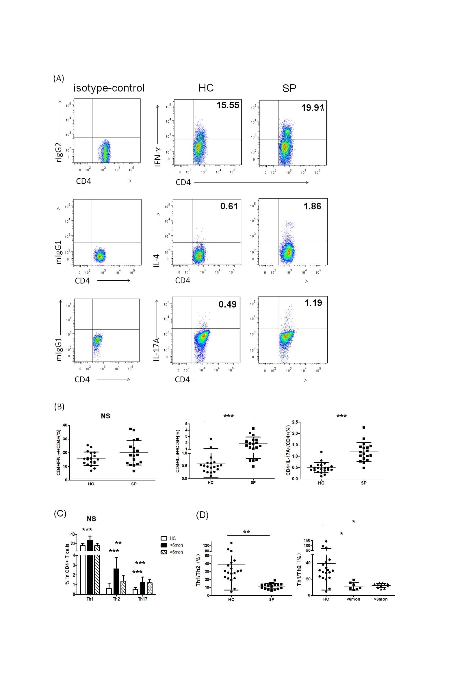
Th2 Biased Immunity With Altered B Cell Profiles in Circulation of Patients With Sporotrichosis Caused by Sporothrix globosa.
Simian-Human Immunodeficiency Virus SHIV.CH505 Infection of Rhesus Macaques Results in Persistent Viral Replication and Induces Intestinal Immunopathology.
Tim-3 regulates inflammatory cytokine expression and Th17 cell response induced by monocytes from patients with chronic hepatitis B.
Th17 and IL-17 exhibit higher levels in osteonecrosis of the femoral head and have a positive correlation with severity of pain.
The Genetic Polymorphisms of NLRP3 Inflammasome Associated with T Helper Cells in Patients with Multiple Myeloma.
DNA Methyltransferase Inhibition Promotes Th1 Polarization in Human CD4(+)CD25(high) FOXP3(+) Regulatory T Cells but Does Not Affect Their Suppressive Capacity.
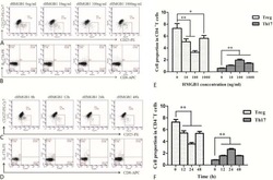
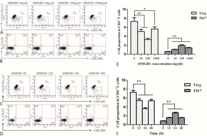
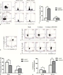
The Effects of High Mobility Group Box-1 Protein on Peripheral Treg/Th17 Balance in Patients with Atherosclerosis.
Intestinal damage precedes mucosal immune dysfunction in SIV infection.
P2X7R mutation disrupts the NLRP3-mediated Th program and predicts poor cardiac allograft outcomes.
Monomeric Immunoglobulin A from Plasma Inhibits Human Th17 Responses In Vitro Independent of FcαRI and DC-SIGN.
Application of a whole blood mycobacterial growth inhibition assay to study immunity against Mycobacterium tuberculosis in a high tuberculosis burden population.
Suppressive IL-17A(+)Foxp3(+) and ex-Th17 IL-17A(neg)Foxp3(+) T(reg) cells are a source of tumour-associated T(reg) cells.
Immune cell-derived cytokines contribute to obesity-related inflammation, fibrogenesis and metabolic deregulation in human adipose tissue.
Human Zika infection induces a reduction of IFN-γ producing CD4 T-cells and a parallel expansion of effector Vδ2 T-cells.
IL-1β, But Not Programed Death-1 and Programed Death Ligand Pathway, Is Critical for the Human Th17 Response to Mycobacterium tuberculosis.
Characterization and Quantification of Innate Lymphoid Cell Subsets in Human Lung.
Downregulation of RUNX3 moderates the frequency of Th17 and Th22 cells in patients with psoriasis.
Equilibrium of Treg/Th17 cells of peripheral blood in syphilitic patients with sero-resistance.
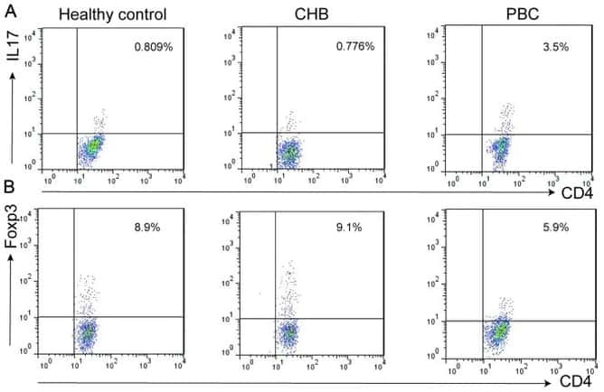
Altered expression of miR-92a correlates with Th17 cell frequency in patients with primary biliary cirrhosis.
Effects of transcutaneous acupoint electrical stimulation on the imbalance of Th(1), Th(2), Th(17) and T(reg) cells following thoracotomy of patients with lung cancer.
Preclinical Testing of Antihuman CD28 Fab' Antibody in a Novel Nonhuman Primate Small Animal Rodent Model of Xenogenic Graft-Versus-Host Disease.
Jejunal T Cell Inflammation in Human Obesity Correlates with Decreased Enterocyte Insulin Signaling.
CARD9-Dependent Neutrophil Recruitment Protects against Fungal Invasion of the Central Nervous System.
Severe H7N9 infection is associated with decreased antigen-presenting capacity of CD14+ cells.
Breast-fed and bottle-fed infant rhesus macaques develop distinct gut microbiotas and immune systems.
CD4+ T cells contain early extrapulmonary tuberculosis (TB) dissemination and rapid TB progression and sustain multieffector functions of CD8+ T and CD3- lymphocytes: mechanisms of CD4+ T cell immunity.
T cell-derived IL-22 amplifies IL-1β-driven inflammation in human adipose tissue: relevance to obesity and type 2 diabetes.
CCL20 Secretion from the Nucleus Pulposus Improves the Recruitment of CCR6-Expressing Th17 Cells to Degenerated IVD Tissues.
Intercellular adhesion molecule 1 mediates migration of Th1 and Th17 cells across human retinal vascular endothelium.
Reconstitution of interleukin-17-producing T helper cells after allogeneic hematopoietic cell transplantation.
Early and nonreversible decrease of CD161++ /MAIT cells in HIV infection.
Reduced frequency of memory T cells and increased Th17 responses in patients with active tuberculosis.
IL-2 simultaneously expands Foxp3+ T regulatory and T effector cells and confers resistance to severe tuberculosis (TB): implicative Treg-T effector cooperation in immunity to TB.
IL-17 and IFN-γ expression in lymphocytes from patients with active tuberculosis correlates with the severity of the disease.
Identification of Mycobacterium tuberculosis-specific Th1, Th17 and Th22 cells using the expression of CD40L in tuberculous pleurisy.
T-cell immune function in tumor, skin, and peripheral blood of advanced stage melanoma patients: implications for immunotherapy.
Human slan (6-sulfo LacNAc) dendritic cells are inflammatory dermal dendritic cells in psoriasis and drive strong TH17/TH1 T-cell responses.
Regulation of IL-17 in chronic inflammation in the human lung.
Human MAIT cells are xenobiotic-resistant, tissue-targeted, CD161hi IL-17-secreting T cells.
Strong viremia control in vaccinated macaques does not prevent gradual Th17 cell loss from central memory.
Specific T cell frequency and cytokine expression profile do not correlate with protection against tuberculosis after bacillus Calmette-Guérin vaccination of newborns.
Mechanisms underlying γδ T-cell subset perturbations in SIV-infected Asian rhesus macaques.
Cytokine requirements for the differentiation and expansion of IL-17A- and IL-22-producing human Vgamma2Vdelta2 T cells.
Compromised gastrointestinal integrity in pigtail macaques is associated with increased microbial translocation, immune activation, and IL-17 production in the absence of SIV infection.
Intensive chemotherapy for acute myeloid leukemia differentially affects circulating TC1, TH1, TH17 and TREG cells.
Differentiation, distribution and gammadelta T cell-driven regulation of IL-22-producing T cells in tuberculosis.
In colorectal cancer mast cells contribute to systemic regulatory T-cell dysfunction.
Autoimmune destruction of skin melanocytes by perilesional T cells from vitiligo patients.
1,25-Dihydroxyvitamin D3 and IL-2 combine to inhibit T cell production of inflammatory cytokines and promote development of regulatory T cells expressing CTLA-4 and FoxP3.
A monoclonal antibody selection for immunohistochemical examination of lymphoid tissues from non-human primates.
Distinct regulation of interleukin-17 in human T helper lymphocytes.
Obasanmi G, Lois N, Armstrong D, Hombrebueno JMR, Lynch A, Chen M, Xu H
Cells 2023 Jan 31;12(3)
Cells 2023 Jan 31;12(3)
MBL Binding with AhR Controls Th17 Immunity in Silicosis-Associated Lung Inflammation and Fibrosis.
Liu Y, Zhao N, Xu Q, Deng F, Wang P, Dong L, Lu X, Xia L, Wang M, Chen Z, Zhou J, Zuo D
Journal of inflammation research 2022;15:4315-4329
Journal of inflammation research 2022;15:4315-4329
Cytomegalovirus mediates expansion of IL-15-responsive innate-memory cells with SIV killing function.
Méndez-Lagares G, Chin N, Chang WLW, Lee J, Rosás-Umbert M, Kieu HT, Merriam D, Lu W, Kim S, Adamson L, Brander C, Luciw PA, Barry PA, Hartigan-O'Connor DJ
The Journal of clinical investigation 2021 Aug 2;131(15)
The Journal of clinical investigation 2021 Aug 2;131(15)
Th2 Biased Immunity With Altered B Cell Profiles in Circulation of Patients With Sporotrichosis Caused by Sporothrix globosa.
Zu J, Yao L, Song Y, Cui Y, Guan M, Chen R, Zhen Y, Li S
Frontiers in immunology 2020;11:570888
Frontiers in immunology 2020;11:570888
Simian-Human Immunodeficiency Virus SHIV.CH505 Infection of Rhesus Macaques Results in Persistent Viral Replication and Induces Intestinal Immunopathology.
Bar KJ, Coronado E, Hensley-McBain T, O'Connor MA, Osborn JM, Miller C, Gott TM, Wangari S, Iwayama N, Ahrens CY, Smedley J, Moats C, Lynch RM, Haddad EK, Haigwood NL, Fuller DH, Shaw GM, Klatt NR, Manuzak JA
Journal of virology 2019 Sep 15;93(18)
Journal of virology 2019 Sep 15;93(18)
Tim-3 regulates inflammatory cytokine expression and Th17 cell response induced by monocytes from patients with chronic hepatitis B.
Wang J, Li C, Fu J, Wang X, Feng X, Pan X
Scandinavian journal of immunology 2019 May;89(5):e12755
Scandinavian journal of immunology 2019 May;89(5):e12755
Th17 and IL-17 exhibit higher levels in osteonecrosis of the femoral head and have a positive correlation with severity of pain.
Zou D, Zhang K, Yang Y, Ren Y, Zhang L, Xiao X, Zhang H, Liu S, Li J
Endokrynologia Polska 2018;69(3):283-290
Endokrynologia Polska 2018;69(3):283-290
The Genetic Polymorphisms of NLRP3 Inflammasome Associated with T Helper Cells in Patients with Multiple Myeloma.
Zhao X, Hua M, Yan S, Yu J, Han F, Zhong C, Wang R, Zhang C, Hou M, Ma D
Journal of immunology research 2018;2018:7569809
Journal of immunology research 2018;2018:7569809
DNA Methyltransferase Inhibition Promotes Th1 Polarization in Human CD4(+)CD25(high) FOXP3(+) Regulatory T Cells but Does Not Affect Their Suppressive Capacity.
Landman S, Cruijsen M, Urbano PCM, Huls G, van Erp PEJ, van Rijssen E, Joosten I, Koenen HJPM
Journal of immunology research 2018;2018:4973964
Journal of immunology research 2018;2018:4973964
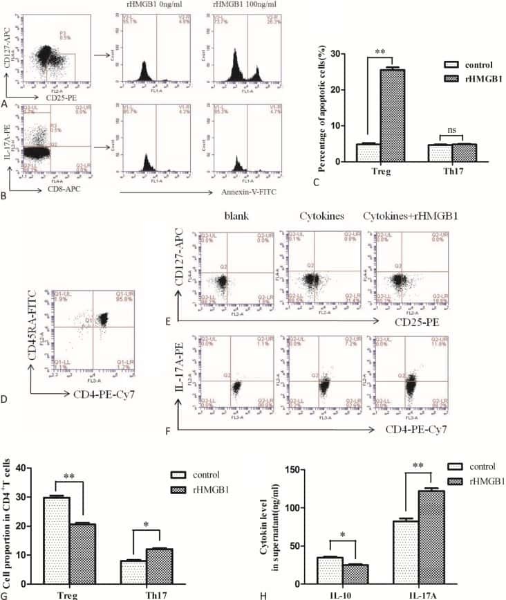
The Effects of High Mobility Group Box-1 Protein on Peripheral Treg/Th17 Balance in Patients with Atherosclerosis.
Ding JW, Zhou T, Zheng XX, Wang XA, Tong XH, Luo CY, Zhang ZQ, Yu B
Acta Cardiologica Sinica 2018 Sep;34(5):399-408
Acta Cardiologica Sinica 2018 Sep;34(5):399-408
Intestinal damage precedes mucosal immune dysfunction in SIV infection.
Hensley-McBain T, Berard AR, Manuzak JA, Miller CJ, Zevin AS, Polacino P, Gile J, Agricola B, Cameron M, Hu SL, Estes JD, Reeves RK, Smedley J, Keele BF, Burgener AD, Klatt NR
Mucosal immunology 2018 Sep;11(5):1429-1440
Mucosal immunology 2018 Sep;11(5):1429-1440
P2X7R mutation disrupts the NLRP3-mediated Th program and predicts poor cardiac allograft outcomes.
D'Addio F, Vergani A, Potena L, Maestroni A, Usuelli V, Ben Nasr M, Bassi R, Tezza S, Dellepiane S, El Essawy B, Iascone M, Iacovoni A, Borgese L, Liu K, Visner G, Dhe-Paganon S, Corradi D, Abdi R, Starling RC, Folli F, Zuccotti GV, Sayegh MH, Heeger PS, Chandraker A, Grigioni F, Fiorina P
The Journal of clinical investigation 2018 Aug 1;128(8):3490-3503
The Journal of clinical investigation 2018 Aug 1;128(8):3490-3503
Monomeric Immunoglobulin A from Plasma Inhibits Human Th17 Responses In Vitro Independent of FcαRI and DC-SIGN.
Saha C, Das M, Patil V, Stephen-Victor E, Sharma M, Wymann S, Jordi M, Vonarburg C, Kaveri SV, Bayry J
Frontiers in immunology 2017;8:275
Frontiers in immunology 2017;8:275
Application of a whole blood mycobacterial growth inhibition assay to study immunity against Mycobacterium tuberculosis in a high tuberculosis burden population.
Baguma R, Penn-Nicholson A, Smit E, Erasmus M, Day J, Makhethe L, de Kock M, Hughes EJ, van Rooyen M, Pienaar B, Stone L, Hanekom W, Brennan MJ, Wallis RS, Hatherill M, Scriba TJ
PloS one 2017;12(9):e0184563
PloS one 2017;12(9):e0184563
Suppressive IL-17A(+)Foxp3(+) and ex-Th17 IL-17A(neg)Foxp3(+) T(reg) cells are a source of tumour-associated T(reg) cells.
Downs-Canner S, Berkey S, Delgoffe GM, Edwards RP, Curiel T, Odunsi K, Bartlett DL, Obermajer N
Nature communications 2017 Mar 14;8:14649
Nature communications 2017 Mar 14;8:14649
Immune cell-derived cytokines contribute to obesity-related inflammation, fibrogenesis and metabolic deregulation in human adipose tissue.
Caër C, Rouault C, Le Roy T, Poitou C, Aron-Wisnewsky J, Torcivia A, Bichet JC, Clément K, Guerre-Millo M, André S
Scientific reports 2017 Jun 7;7(1):3000
Scientific reports 2017 Jun 7;7(1):3000
Human Zika infection induces a reduction of IFN-γ producing CD4 T-cells and a parallel expansion of effector Vδ2 T-cells.
Cimini E, Castilletti C, Sacchi A, Casetti R, Bordoni V, Romanelli A, Turchi F, Martini F, Tumino N, Nicastri E, Corpolongo A, Di Caro A, Kobinger G, Zumla A, Capobianchi MR, Ippolito G, Agrati C
Scientific reports 2017 Jul 24;7(1):6313
Scientific reports 2017 Jul 24;7(1):6313
IL-1β, But Not Programed Death-1 and Programed Death Ligand Pathway, Is Critical for the Human Th17 Response to Mycobacterium tuberculosis.
Stephen-Victor E, Sharma VK, Das M, Karnam A, Saha C, Lecerf M, Galeotti C, Kaveri SV, Bayry J
Frontiers in immunology 2016;7:465
Frontiers in immunology 2016;7:465
Characterization and Quantification of Innate Lymphoid Cell Subsets in Human Lung.
De Grove KC, Provoost S, Verhamme FM, Bracke KR, Joos GF, Maes T, Brusselle GG
PloS one 2016;11(1):e0145961
PloS one 2016;11(1):e0145961
Downregulation of RUNX3 moderates the frequency of Th17 and Th22 cells in patients with psoriasis.
Fu D, Song X, Hu H, Sun M, Li Z, Tian Z
Molecular medicine reports 2016 Jun;13(6):4606-12
Molecular medicine reports 2016 Jun;13(6):4606-12
Equilibrium of Treg/Th17 cells of peripheral blood in syphilitic patients with sero-resistance.
Zhao J, Ma J, Zhang X, Li Q, Yang X
Experimental and therapeutic medicine 2016 Jun;11(6):2300-2304
Experimental and therapeutic medicine 2016 Jun;11(6):2300-2304
Altered expression of miR-92a correlates with Th17 cell frequency in patients with primary biliary cirrhosis.
Liang DY, Hou YQ, Luo LJ, Ao L
International journal of molecular medicine 2016 Jul;38(1):131-8
International journal of molecular medicine 2016 Jul;38(1):131-8
Effects of transcutaneous acupoint electrical stimulation on the imbalance of Th(1), Th(2), Th(17) and T(reg) cells following thoracotomy of patients with lung cancer.
Wu H, Wang K, Li G, Meng D, Han J, Wang G, Li YU
Experimental and therapeutic medicine 2016 Feb;11(2):495-502
Experimental and therapeutic medicine 2016 Feb;11(2):495-502
Preclinical Testing of Antihuman CD28 Fab' Antibody in a Novel Nonhuman Primate Small Animal Rodent Model of Xenogenic Graft-Versus-Host Disease.
Hippen KL, Watkins B, Tkachev V, Lemire AM, Lehnen C, Riddle MJ, Singh K, Panoskaltsis-Mortari A, Vanhove B, Tolar J, Kean LS, Blazar BR
Transplantation 2016 Dec;100(12):2630-2639
Transplantation 2016 Dec;100(12):2630-2639
Jejunal T Cell Inflammation in Human Obesity Correlates with Decreased Enterocyte Insulin Signaling.
Monteiro-Sepulveda M, Touch S, Mendes-Sá C, André S, Poitou C, Allatif O, Cotillard A, Fohrer-Ting H, Hubert EL, Remark R, Genser L, Tordjman J, Garbin K, Osinski C, Sautès-Fridman C, Leturque A, Clément K, Brot-Laroche E
Cell metabolism 2015 Jul 7;22(1):113-24
Cell metabolism 2015 Jul 7;22(1):113-24
CARD9-Dependent Neutrophil Recruitment Protects against Fungal Invasion of the Central Nervous System.
Drummond RA, Collar AL, Swamydas M, Rodriguez CA, Lim JK, Mendez LM, Fink DL, Hsu AP, Zhai B, Karauzum H, Mikelis CM, Rose SR, Ferre EM, Yockey L, Lemberg K, Kuehn HS, Rosenzweig SD, Lin X, Chittiboina P, Datta SK, Belhorn TH, Weimer ET, Hernandez ML, Hohl TM, Kuhns DB, Lionakis MS
PLoS pathogens 2015 Dec;11(12):e1005293
PLoS pathogens 2015 Dec;11(12):e1005293
Severe H7N9 infection is associated with decreased antigen-presenting capacity of CD14+ cells.
Diao H, Cui G, Wei Y, Chen J, Zuo J, Cao H, Chen Y, Yao H, Tian Z, Li L
PloS one 2014;9(3):e92823
PloS one 2014;9(3):e92823
Breast-fed and bottle-fed infant rhesus macaques develop distinct gut microbiotas and immune systems.
Ardeshir A, Narayan NR, Méndez-Lagares G, Lu D, Rauch M, Huang Y, Van Rompay KK, Lynch SV, Hartigan-O'Connor DJ
Science translational medicine 2014 Sep 3;6(252):252ra120
Science translational medicine 2014 Sep 3;6(252):252ra120
CD4+ T cells contain early extrapulmonary tuberculosis (TB) dissemination and rapid TB progression and sustain multieffector functions of CD8+ T and CD3- lymphocytes: mechanisms of CD4+ T cell immunity.
Yao S, Huang D, Chen CY, Halliday L, Wang RC, Chen ZW
Journal of immunology (Baltimore, Md. : 1950) 2014 Mar 1;192(5):2120-32
Journal of immunology (Baltimore, Md. : 1950) 2014 Mar 1;192(5):2120-32
T cell-derived IL-22 amplifies IL-1β-driven inflammation in human adipose tissue: relevance to obesity and type 2 diabetes.
Dalmas E, Venteclef N, Caer C, Poitou C, Cremer I, Aron-Wisnewsky J, Lacroix-Desmazes S, Bayry J, Kaveri SV, Clément K, André S, Guerre-Millo M
Diabetes 2014 Jun;63(6):1966-77
Diabetes 2014 Jun;63(6):1966-77
CCL20 Secretion from the Nucleus Pulposus Improves the Recruitment of CCR6-Expressing Th17 Cells to Degenerated IVD Tissues.
Zhang W, Nie L, Wang Y, Wang XP, Zhao H, Dongol S, Maharjan S, Cheng L
PloS one 2013;8(6):e66286
PloS one 2013;8(6):e66286
Intercellular adhesion molecule 1 mediates migration of Th1 and Th17 cells across human retinal vascular endothelium.
Bharadwaj AS, Schewitz-Bowers LP, Wei L, Lee RW, Smith JR
Investigative ophthalmology & visual science 2013 Oct 23;54(10):6917-25
Investigative ophthalmology & visual science 2013 Oct 23;54(10):6917-25
Reconstitution of interleukin-17-producing T helper cells after allogeneic hematopoietic cell transplantation.
Bahr F, Wehner R, Platzbecker U, Wermke M, Shayegi N, Middeke JM, Röllig C, Schetelig J, Ehninger G, Schmitz M, Bornhäuser M, Tuve S
Biology of blood and marrow transplantation : journal of the American Society for Blood and Marrow Transplantation 2013 Mar;19(3):357-65
Biology of blood and marrow transplantation : journal of the American Society for Blood and Marrow Transplantation 2013 Mar;19(3):357-65
Early and nonreversible decrease of CD161++ /MAIT cells in HIV infection.
Cosgrove C, Ussher JE, Rauch A, Gärtner K, Kurioka A, Hühn MH, Adelmann K, Kang YH, Fergusson JR, Simmonds P, Goulder P, Hansen TH, Fox J, Günthard HF, Khanna N, Powrie F, Steel A, Gazzard B, Phillips RE, Frater J, Uhlig H, Klenerman P
Blood 2013 Feb 7;121(6):951-61
Blood 2013 Feb 7;121(6):951-61
Reduced frequency of memory T cells and increased Th17 responses in patients with active tuberculosis.
Marín ND, París SC, Rojas M, García LF
Clinical and vaccine immunology : CVI 2012 Oct;19(10):1667-76
Clinical and vaccine immunology : CVI 2012 Oct;19(10):1667-76
IL-2 simultaneously expands Foxp3+ T regulatory and T effector cells and confers resistance to severe tuberculosis (TB): implicative Treg-T effector cooperation in immunity to TB.
Chen CY, Huang D, Yao S, Halliday L, Zeng G, Wang RC, Chen ZW
Journal of immunology (Baltimore, Md. : 1950) 2012 May 1;188(9):4278-88
Journal of immunology (Baltimore, Md. : 1950) 2012 May 1;188(9):4278-88
IL-17 and IFN-γ expression in lymphocytes from patients with active tuberculosis correlates with the severity of the disease.
Jurado JO, Pasquinelli V, Alvarez IB, Peña D, Rovetta AI, Tateosian NL, Romeo HE, Musella RM, Palmero D, Chuluyán HE, García VE
Journal of leukocyte biology 2012 Jun;91(6):991-1002
Journal of leukocyte biology 2012 Jun;91(6):991-1002
Identification of Mycobacterium tuberculosis-specific Th1, Th17 and Th22 cells using the expression of CD40L in tuberculous pleurisy.
Li L, Qiao D, Fu X, Lao S, Zhang X, Wu C
PloS one 2011;6(5):e20165
PloS one 2011;6(5):e20165
T-cell immune function in tumor, skin, and peripheral blood of advanced stage melanoma patients: implications for immunotherapy.
Tjin EP, Konijnenberg D, Krebbers G, Mallo H, Drijfhout JW, Franken KL, van der Horst CM, Bos JD, Nieweg OE, Kroon BB, Haanen JB, Melief CJ, Vyth-Dreese FA, Luiten RM
Clinical cancer research : an official journal of the American Association for Cancer Research 2011 Sep 1;17(17):5736-47
Clinical cancer research : an official journal of the American Association for Cancer Research 2011 Sep 1;17(17):5736-47
Human slan (6-sulfo LacNAc) dendritic cells are inflammatory dermal dendritic cells in psoriasis and drive strong TH17/TH1 T-cell responses.
Hänsel A, Günther C, Ingwersen J, Starke J, Schmitz M, Bachmann M, Meurer M, Rieber EP, Schäkel K
The Journal of allergy and clinical immunology 2011 Mar;127(3):787-94.e1-9
The Journal of allergy and clinical immunology 2011 Mar;127(3):787-94.e1-9
Regulation of IL-17 in chronic inflammation in the human lung.
Pridgeon C, Bugeon L, Donnelly L, Straschil U, Tudhope SJ, Fenwick P, Lamb JR, Barnes PJ, Dallman MJ
Clinical science (London, England : 1979) 2011 Jun;120(12):515-24
Clinical science (London, England : 1979) 2011 Jun;120(12):515-24
Human MAIT cells are xenobiotic-resistant, tissue-targeted, CD161hi IL-17-secreting T cells.
Dusseaux M, Martin E, Serriari N, Péguillet I, Premel V, Louis D, Milder M, Le Bourhis L, Soudais C, Treiner E, Lantz O
Blood 2011 Jan 27;117(4):1250-9
Blood 2011 Jan 27;117(4):1250-9
Strong viremia control in vaccinated macaques does not prevent gradual Th17 cell loss from central memory.
Demberg T, Ettinger AC, Aladi S, McKinnon K, Kuddo T, Venzon D, Patterson LJ, Phillips TM, Robert-Guroff M
Vaccine 2011 Aug 11;29(35):6017-28
Vaccine 2011 Aug 11;29(35):6017-28
Specific T cell frequency and cytokine expression profile do not correlate with protection against tuberculosis after bacillus Calmette-Guérin vaccination of newborns.
Kagina BM, Abel B, Scriba TJ, Hughes EJ, Keyser A, Soares A, Gamieldien H, Sidibana M, Hatherill M, Gelderbloem S, Mahomed H, Hawkridge A, Hussey G, Kaplan G, Hanekom WA, other members of the South African Tuberculosis Vaccine Initiative
American journal of respiratory and critical care medicine 2010 Oct 15;182(8):1073-9
American journal of respiratory and critical care medicine 2010 Oct 15;182(8):1073-9
Mechanisms underlying γδ T-cell subset perturbations in SIV-infected Asian rhesus macaques.
Harris LD, Klatt NR, Vinton C, Briant JA, Tabb B, Ladell K, Lifson J, Estes JD, Price DA, Hirsch VM, Brenchley JM
Blood 2010 Nov 18;116(20):4148-57
Blood 2010 Nov 18;116(20):4148-57
Cytokine requirements for the differentiation and expansion of IL-17A- and IL-22-producing human Vgamma2Vdelta2 T cells.
Ness-Schwickerath KJ, Jin C, Morita CT
Journal of immunology (Baltimore, Md. : 1950) 2010 Jun 15;184(12):7268-80
Journal of immunology (Baltimore, Md. : 1950) 2010 Jun 15;184(12):7268-80
Compromised gastrointestinal integrity in pigtail macaques is associated with increased microbial translocation, immune activation, and IL-17 production in the absence of SIV infection.
Klatt NR, Harris LD, Vinton CL, Sung H, Briant JA, Tabb B, Morcock D, McGinty JW, Lifson JD, Lafont BA, Martin MA, Levine AD, Estes JD, Brenchley JM
Mucosal immunology 2010 Jul;3(4):387-98
Mucosal immunology 2010 Jul;3(4):387-98
Intensive chemotherapy for acute myeloid leukemia differentially affects circulating TC1, TH1, TH17 and TREG cells.
Ersvaer E, Liseth K, Skavland J, Gjertsen BT, Bruserud Ø
BMC immunology 2010 Jul 9;11:38
BMC immunology 2010 Jul 9;11:38
Differentiation, distribution and gammadelta T cell-driven regulation of IL-22-producing T cells in tuberculosis.
Yao S, Huang D, Chen CY, Halliday L, Zeng G, Wang RC, Chen ZW
PLoS pathogens 2010 Feb 26;6(2):e1000789
PLoS pathogens 2010 Feb 26;6(2):e1000789
In colorectal cancer mast cells contribute to systemic regulatory T-cell dysfunction.
Blatner NR, Bonertz A, Beckhove P, Cheon EC, Krantz SB, Strouch M, Weitz J, Koch M, Halverson AL, Bentrem DJ, Khazaie K
Proceedings of the National Academy of Sciences of the United States of America 2010 Apr 6;107(14):6430-5
Proceedings of the National Academy of Sciences of the United States of America 2010 Apr 6;107(14):6430-5
Autoimmune destruction of skin melanocytes by perilesional T cells from vitiligo patients.
van den Boorn JG, Konijnenberg D, Dellemijn TA, van der Veen JP, Bos JD, Melief CJ, Vyth-Dreese FA, Luiten RM
The Journal of investigative dermatology 2009 Sep;129(9):2220-32
The Journal of investigative dermatology 2009 Sep;129(9):2220-32
1,25-Dihydroxyvitamin D3 and IL-2 combine to inhibit T cell production of inflammatory cytokines and promote development of regulatory T cells expressing CTLA-4 and FoxP3.
Jeffery LE, Burke F, Mura M, Zheng Y, Qureshi OS, Hewison M, Walker LS, Lammas DA, Raza K, Sansom DM
Journal of immunology (Baltimore, Md. : 1950) 2009 Nov 1;183(9):5458-67
Journal of immunology (Baltimore, Md. : 1950) 2009 Nov 1;183(9):5458-67
A monoclonal antibody selection for immunohistochemical examination of lymphoid tissues from non-human primates.
Kap YS, van Meurs M, van Driel N, Koopman G, Melief MJ, Brok HP, Laman JD, 't Hart BA
The journal of histochemistry and cytochemistry : official journal of the Histochemistry Society 2009 Dec;57(12):1159-67
The journal of histochemistry and cytochemistry : official journal of the Histochemistry Society 2009 Dec;57(12):1159-67
Distinct regulation of interleukin-17 in human T helper lymphocytes.
Chen Z, Tato CM, Muul L, Laurence A, O'Shea JJ
Arthritis and rheumatism 2007 Sep;56(9):2936-46
Arthritis and rheumatism 2007 Sep;56(9):2936-46
No comments: Submit comment
Supportive validation
- Submitted by
- Invitrogen Antibodies (provider)
- Main image
- Experimental details
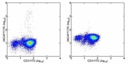
- Normal human peripheral blood cells were stimulated overnight with PMA and Ionomycin in the presence of monensin. Cells were stained with Anti-Human CD3 FITC (Product # 11-0037-42), then fixed, permeablized, and stained with Anti-Human IL-17A PE (Left figure). The ligand blocking control demonstrates that pre-blocking Anti-Human IL-17A PE with 1.0 µg of Human IL-17A Recombinant Protein (Product # 14-8179-80) prior to staining abrogates specific IL-17A staining (Right figure). Cells in the lymphocyte gate were used for analysis.
- Conjugate
- Yellow dye

Supportive validation
- Submitted by
- Invitrogen Antibodies (provider)
- Main image

- Experimental details
- NULL
- Conjugate
- Yellow dye
- Submitted by
- Invitrogen Antibodies (provider)
- Main image
- Experimental details
- NULL
- Conjugate
- Yellow dye
- Submitted by
- Invitrogen Antibodies (provider)
- Main image

- Experimental details
- NULL
- Conjugate
- Yellow dye

- Submitted by
- Invitrogen Antibodies (provider)
- Main image
- Experimental details
- NULL
- Conjugate
- Yellow dye
- Submitted by
- Invitrogen Antibodies (provider)
- Main image
- Experimental details
- NULL
- Conjugate
- Yellow dye
- Submitted by
- Invitrogen Antibodies (provider)
- Main image
- Experimental details
- NULL
- Conjugate
- Yellow dye
- Submitted by
- Invitrogen Antibodies (provider)
- Main image
- Experimental details
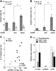
- Figure 1 Obese omental adipose tissue microenvironment promotes IL-17 release. ( A ) IL-17 + and IL-22 + cells were quantified by FACS analysis of blood-derived memory CD4 + T cells cultured for 7 days without or with conditioned medium of omental adipose tissue (omCM) from lean (n = 3) or obese (n = 8) subjects. *P < 0.05. ( B ) IL-17 and IL-22 concentrations were determined by Luminex in blood-derived memory CD4 + T cells culture medium. *P < 0.05. ( C ) Correlation analysis between IL-17 concentration in blood CD4 + T cells culture medium and IL-1beta concentration in omCM. The correlation coefficient (r) and p value were obtained by Spearman's test. (D) IL17RA, IL22RA1 and IL1R1 mRNA were determined in pre-adipocytes (n = 7), CD31 + endothelial cells (n = 6) and adipocytes (n = 4) obtained from lipoaspirate adipose tissue samples. Data are shown as mean +- SEM. nd: not detected.
- Conjugate
- Yellow dye

- Submitted by
- Invitrogen Antibodies (provider)
- Main image

- Experimental details
- Fig 5 Representative flow cytometry plots of cytokine expression by CD4 T cells. CD4 T cells expressing IFN-gamma, IL-2, TNF-alpha and/or IL-17 upon stimulation with BCG or ESAT-6/CFP-10 peptide pools for 12 hours, compared to an unstimulated control sample. The plots represent cytokine-positive cells (red) overlayed onto the background of the entire CD4 T cell parent population (black). Similar assessment was also performed for CD8 and gammadelta T cells (data not shown).
- Conjugate
- Yellow dye

- Submitted by
- Invitrogen Antibodies (provider)
- Main image
- Experimental details
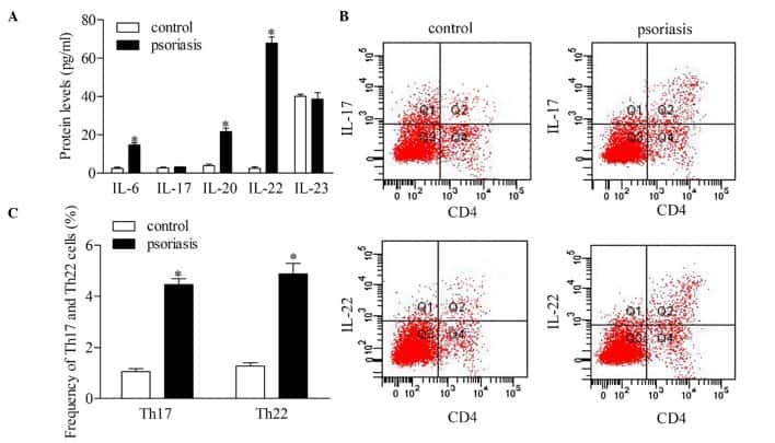
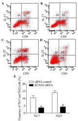
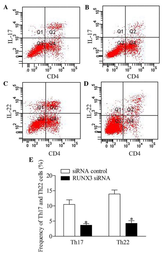
- Figure 5 Inhibition of RUNX3 regulates the frequencies of Th17 and Th22 in CD4 + T cells from patients with psoriasis. The frequencies of Th17 and Th22 cells in CD4 + T cells from patients with psoriasis transfected with RUNX3 siRNA or an siRNA control was detected using flow cytometry. (A and B) Flow cytometric analysis of Th17 in CD4 + T cells from patients with psoriasis. The percentages of cells in the Q2 region represent the percentage of Th17 cells. (C and D) Flow cytometric analysis of Th22 in CD4 + T cells from patients with psoriasis. The percentages of cells in the Q2 region represent the percentage of Th22 cells. (E) Percentages of Th17 and Th22 cells in CD4 + T cells from patients with psoriasis transfected with RUNX3 siRNA or an siRNA control. Data are presented as the mean +- standard deviation of three experiments. * P
- Conjugate
- Yellow dye
- Submitted by
- Invitrogen Antibodies (provider)
- Main image

- Experimental details
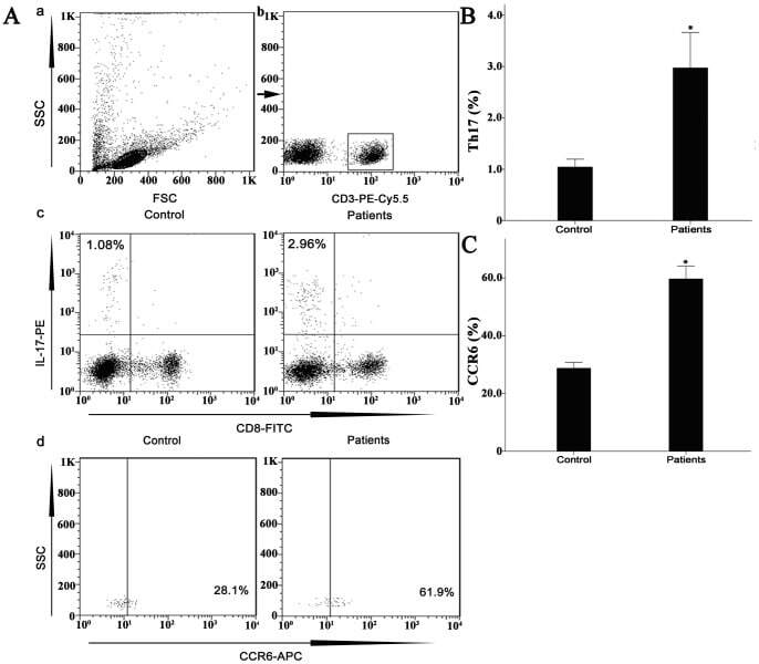
- Figure 7 Circulating percentages of Th17 cells and CCR6-positive cells in peripheral blood are increased in IVD degenerated patients when compared with controls. Heparinized peripheral whole blood cells from 20 patients and 15 healthy controls were stimulated with phorbol myristate acetate (PMA), ionomycin, and monensin for 4 h and subsequently stained with fluorochrome-labeled antibodies as described in Materials and Methods. A(a) Lymphocytes were gated by flow cytometry. A(b) CD3 + T subsets were gated by flow cytometry; the plots in the inset box represent the CD3 + T cells. A(c) Representative IL-17 expression levels in the CD3 + CD8 - T subsets (CD4 + T subsets) from each group are shown. The percentages of positive cells are shown in the upper left panels. A(d) Representative surface CCR6 expression levels on the CD3 + CD8 - IL-17 + subsets from each group are shown. The percentages of positive cells are shown in the right panel. (B) The percentage of circulating Th17 cells was significantly higher in IVD degenerated patients (2.973+-0.689%) than in the control group (1.039+-0.156%; *, p
- Conjugate
- Yellow dye