Explore
 Validate
 Learn
Learn Western blot
 ELISA
ELISAAntibody data
- Antibody Data
- Antigen structure
- References [2]
- Comments [0]
- Validations
- Western blot [1]
Submit
Validation data
Reference
Comment
Report error
- Product number
- NBP1-42746 - Provider product page

- Provider
- Novus Biologicals
- Proper citation
- Novus Cat#NBP1-42746, RRID:AB_10005912
- Product name
- Rabbit Polyclonal IL-17/IL-17A Antibody
- Antibody type
- Polyclonal
- Description
- Immunogen affinity purified. In ELISA and other immunoreactive assays, this antibody will recognize both native and recombinant rat IL17-A in cell supernatants and certain body fluids. A control of similarly diluted normal rabbit IgG is recommended.
- Reactivity
- Human, Mouse, Rat
- Host
- Rabbit
- Conjugate
- Biotin
- Isotype
- IgG
- Vial size
- 0.1 mg
- Concentration
- LYOPH
- Storage
- Store lyophilized antibody at 4C. Aliquot reconstituted liquid and store at -20C. Avoid freeze-thaw cycles.
Submitted references Expression and location of IL-17A, E, F and their receptors in colorectal adenocarcinoma: Comparison with benign intestinal disease.
Expression of IL-17A, E, and F and their receptors in human prostatic cancer: Comparison with benign prostatic hyperplasia.
Liu Y, Sun X, Zhao X, An L, Wang Z, Jiang J, Shen W, Yang X, Sun Y
Pathology, research and practice 2018 Apr;214(4):482-491
Pathology, research and practice 2018 Apr;214(4):482-491
Expression of IL-17A, E, and F and their receptors in human prostatic cancer: Comparison with benign prostatic hyperplasia.
Liu Y, Zhao X, Sun X, Li Y, Wang Z, Jiang J, Han H, Shen W, Corrigan CJ, Sun Y
The Prostate 2015 Dec;75(16):1844-56
The Prostate 2015 Dec;75(16):1844-56
No comments: Submit comment
Supportive validation
- Submitted by
- Novus Biologicals (provider)
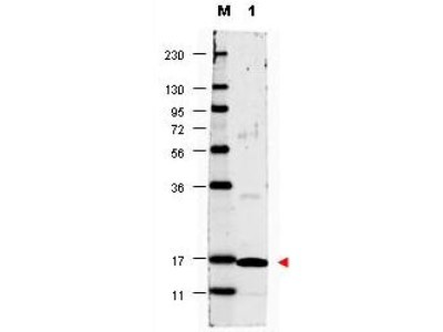
- Main image

- Experimental details
- Western Blot: IL-17/IL-17A Antibody [Biotin] [NBP1-42746] - Analysis of a band 17 kDa in size corresponding to recombinant human IL17-A (lane 1). Molecular weight markers are also shown (M). After transfer, the membrane was blocked overnight with 3% BSA in TBS followed by reaction with primary antibody at a 1:1,000 dilution. Detection occurred using DyLight 649 conjugated anti-Rabbit IgG secondary antibody diluted 1:20,000 in blocking buffer.