Explore
 Validate
 Learn
Learn Immunocytochemistry
Antibody data
- Antibody Data
- Antigen structure
- References [0]
- Comments [0]
- Validations
- Immunocytochemistry [1]
- Immunohistochemistry [1]
- Flow cytometry [3]
Submit
Validation data
Reference
Comment
Report error
- Product number
- 740021M20UG - Provider product page

- Provider
- Invitrogen Antibodies
- Product name
- IL-17A Recombinant Mouse Monoclonal Antibody (eBio64DEC17)
- Antibody type
- Monoclonal
- Antigen
- Other
- Description
- Recombinant Mouse monoclonal antibodies are produced using in vitro expression systems. Recombinant antibodies are produced using specific genes that code for the desired antibodies. These genes are cloned into an expression vector and expressed in vitro. The advantages of recombinant antibodies include better specificity and lot-to-lot consistency. It is recommended that the antibody be carefully titrated for optimal performance in the assay of interest.
- Reactivity
- Human, Mouse
- Host
- Mouse
- Isotype
- IgG
- Antibody clone number
- eBio64DEC17
- Vial size
- 20 µL
- Concentration
- 1.0 mg/mL
- Storage
- Store at 4°C short term. For long term storage, store at -20°C, avoiding freeze/thaw cycles.
No comments: Submit comment
Supportive validation
- Submitted by
- Invitrogen Antibodies (provider)
- Main image
- Experimental details
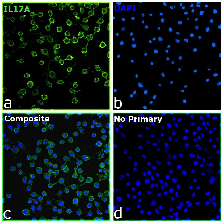
- Immunofluorescent analysis of IL-17A was performed using 70% confluent Neuro-2A cells. The cells were fixed with 4% paraformaldehyde for 10 minutes, permeabilized with 0.1% Triton X-100 for 15 minutes, and blocked with 2% BSA for 1 hour at room temperature. The cells were stained with IL-17A Recombinant Mouse Monoclonal Antibody (eBio64DEC17) (Product # 740021M, 1:100) at 4 degree Celsius overnight and then labeled with Goat anti-Mouse IgG (H+L) Secondary Antibody, Alexa Fluor™ Plus 488 (Product # A32723) at a dilution of 1:2000 for 1 hour at room temperature. Panels a) shows representative image of cells that were stained for detection and localization of IL-17A (green). Panel b) shows representative image of cells stained for nuclei, using DAPI (Product # D1306, 1:500). Panel c) represents a composite image of panels a and b, clearly demonstrating cytoplasmic localization of IL17A. Panel d) represents no primary antibody control to assess the background. The images were captured on EVOS™ M7000 Imaging System (Product # AMF7000) at 20X magnification and externally deconvoluted.
Supportive validation
- Submitted by
- Invitrogen Antibodies (provider)
- Main image
- Experimental details
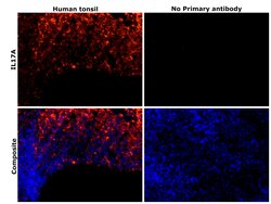
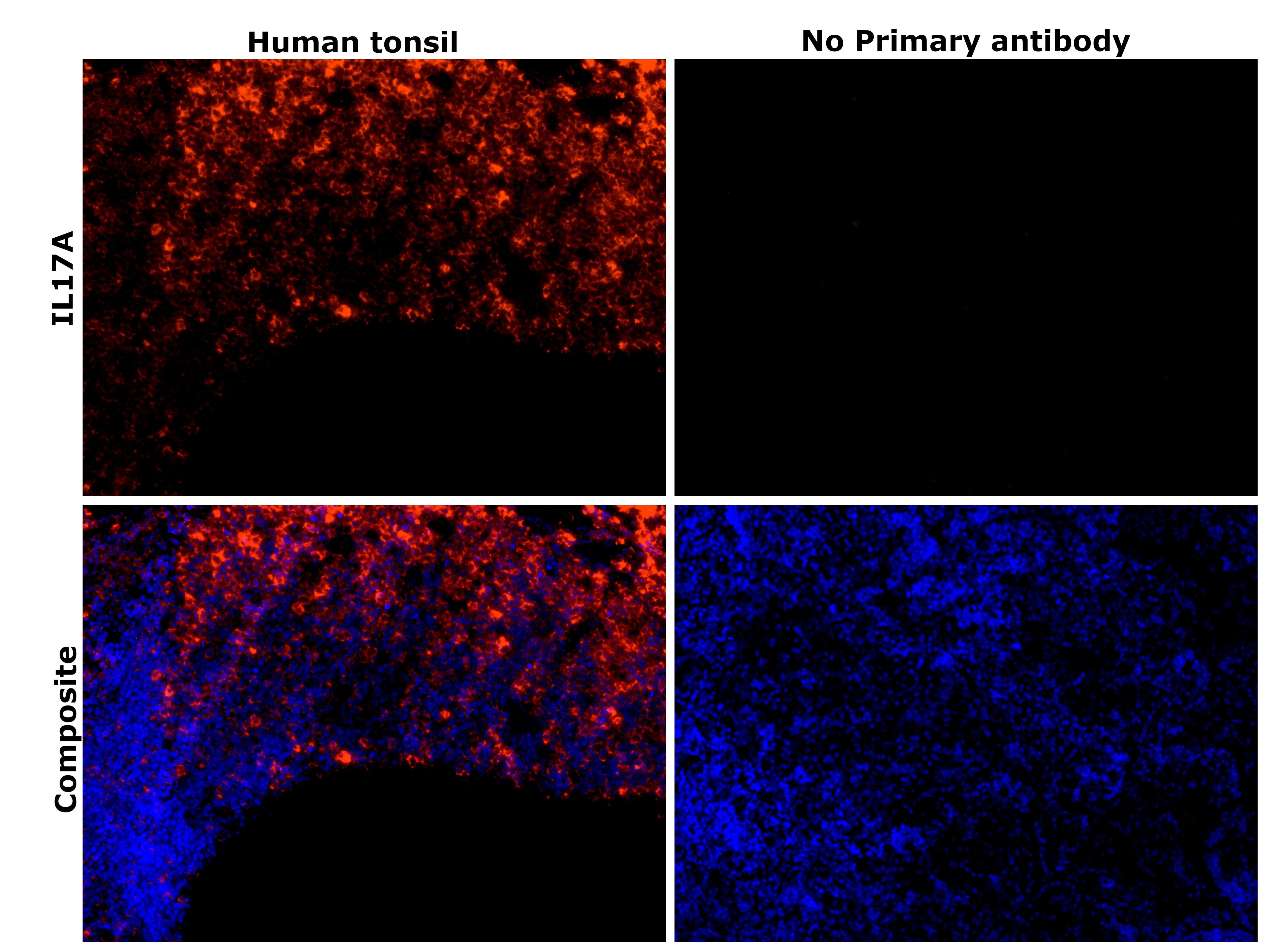
- Immunohistochemical analysis of IL-17A was performed using formalin-fixed paraffin-embedded human tonsil tissue sections. To expose the target protein, heat-induced epitope retrieval was performed on de-paraffinized sections using eBioscience™ IHC Antigen Retrieval Solution - High pH (10X) (Product # 00-4956-58) diluted to 1X solution in water in a decloaking chamber at 110 degree Celsius for 15 minutes. Following antigen retrieval, the sections were blocked with 3% H2O2 for 1h at room temperature followed by 2% normal goat serum in 1X PBS for 45 minutes at room temperature and then probed with or without IL-17A Recombinant Mouse Monoclonal Antibody (eBio64DEC17) (Product # 740021M) at 1:500 dilution in 0.1% normal goat serum overnight at 4 degree Celsius in a humidified chamber. Detection was performed using Alexa Fluor™ 647 Tyramide SuperBoost™ Kit, goat anti-mouse IgG (Product # B40916). Nuclei were stained with DAPI (Product # D1306) and the sections were mounted using ProLong™ Glass Antifade Mountant (Product # P36984). The images were captured on EVOS™ M7000 Imaging System (Product # AMF7000) at 20X magnification.
Supportive validation
- Submitted by
- Invitrogen Antibodies (provider)
- Main image
- Experimental details
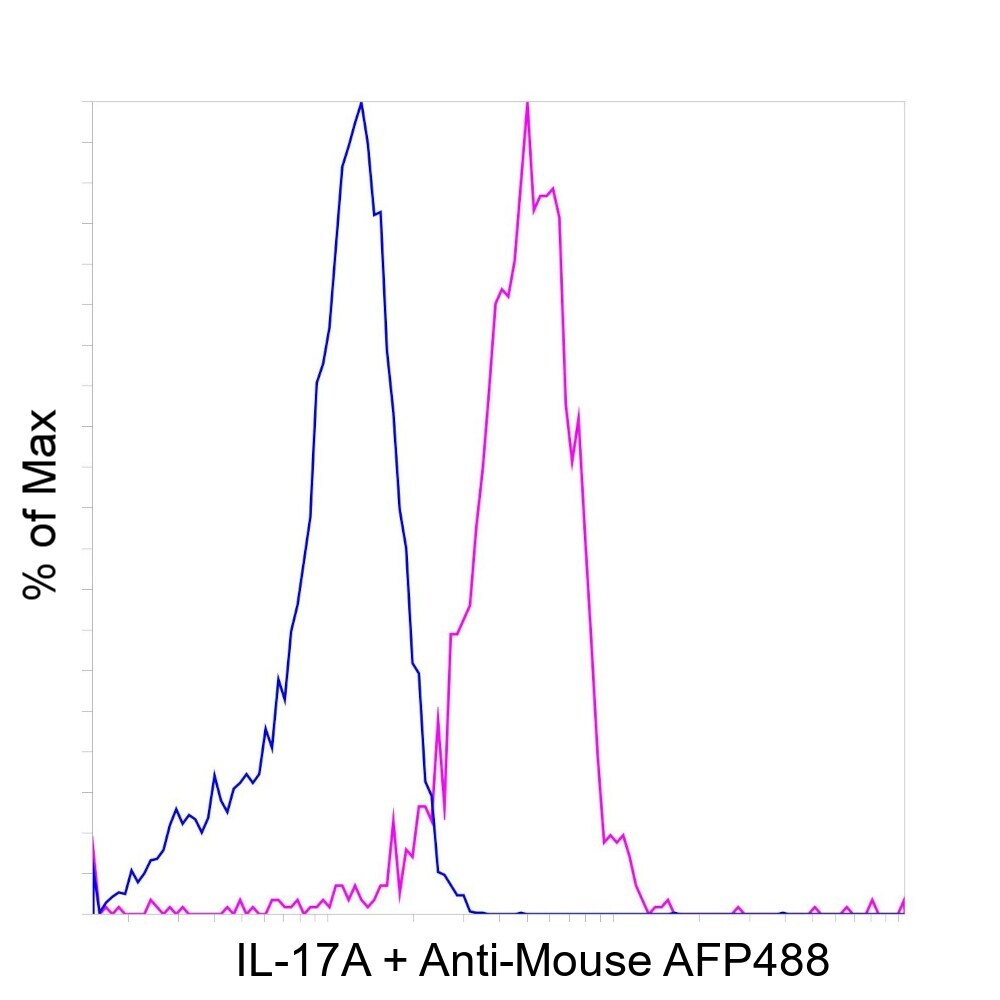
- HEK-293E cells were transiently transfected with human IL-17A construct (pink histogram) or left untransfected (blue histogram). Cells were fixed and permeabilized using the Foxp3 / Transcription Factor Staining Buffer Set (Product # 00-5523-00) and then stained intracellularly with 0.25 µg/test of IL-17A Recombinant Mouse Monoclonal Antibody (eBio64DEC17) (Product # 740021M) (pink histogram) or left untransfected (blue histogram), followed by Goat anti-Mouse IgG (H+L) Alexa Fluor™ Plus 488 (Product # A55058). Viable cells were used for analysis as determined by Fixable Viability Dye eFluor™ 780 (Product # 65-0865-14). The flow cytometry data was acquired using Attune™ NxT Flow Cytometer (Product # A29004).

- Submitted by
- Invitrogen Antibodies (provider)
- Main image
- Experimental details
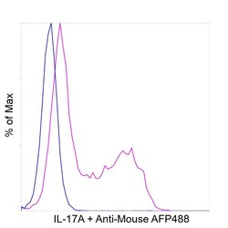
- Flow cytometry analysis of IL-17A was performed using EL4 cells and EL4 cells treated with PMA (50ng/ml) and Ionomycin (500ng/ml) for 6 hours, then with Brefeldin A (500ng/ml) for 18 hours. The cells were fixed and permeabilized using the Foxp3 / Transcription Factor Staining Buffer Set (Product # 00-5523-00) and then stained intracellularly with 2 µg/test of IL-17A Recombinant Mouse Monoclonal Antibody (eBio64DEC17) (Product # 740021M), followed by Goat anti-Mouse IgG (H+L), Alexa Fluor™ Plus 488 (Product # A55058). An increased expression of IL-17A was observed in treated cells (pink histogram) compared to untreated cells (blue histogram). Viable cells were used for analysis, as determined by LIVE/DEAD™ Fixable Violet Dead Cell Stain Kit (Product # L34964). The flow cytometry data was acquired using Attune™ NxT Flow Cytometer (Product # A29004).
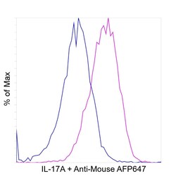
- Submitted by
- Invitrogen Antibodies (provider)
- Main image
- Experimental details
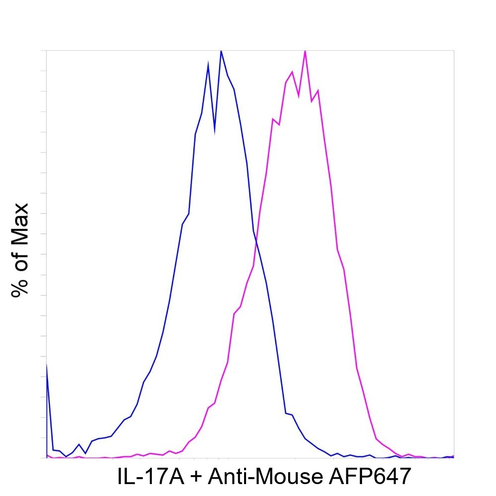
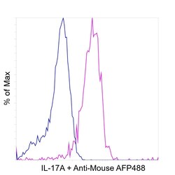
- A549 cells were fixed and permeabilized using the Foxp3 / Transcription Factor Staining Buffer Set (Product # 00-5523-00) and then stained intracellularly with 0.5 µg/test of IL-17A Recombinant Mouse Monoclonal Antibody (eBio64DEC17) (Product # 740021M) (pink histogram) or 0.5 µg/test of Mouse IgG1 K Isotype Control (Product # 14-4714-85) (blue histogram), followed by Goat anti-Mouse IgG (H+L) Alexa Fluor™ Plus 647 (Product # A55060). Viable cells were used for analysis, as determined by LIVE/DEAD™ Fixable Violet Dead Cell Stain Kit (Product # L34964). The flow cytometry data was acquired using Attune™ NxT Flow Cytometer (Product # A29004).